. 2021 Mar 19;5(1):25.
doi: 10.1038/s41698-021-00161-8.

Circulating stromal cells in resectable pancreatic cancer correlates to pathological stage and predicts for poor clinical outcomes

Affiliations

Circulating stromal cells in resectable pancreatic cancer correlates to pathological stage and predicts for poor clinical outcomes

Kirby P Gardner et al. NPJ Precis Oncol. .

Abstract

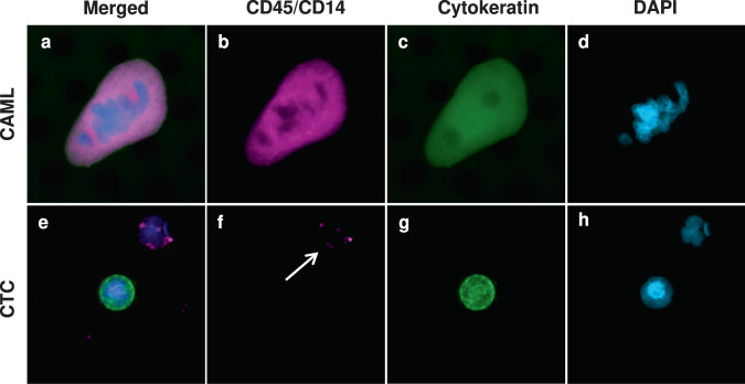
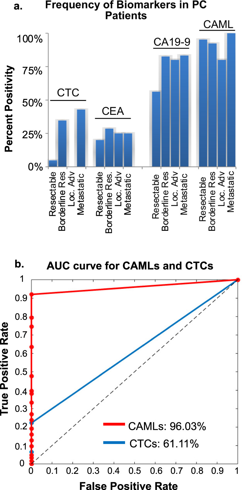
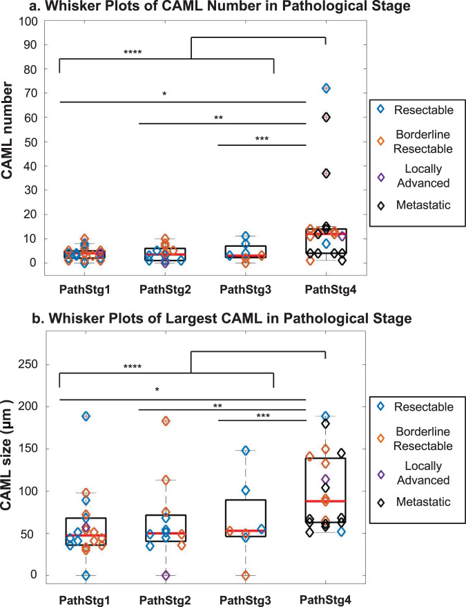
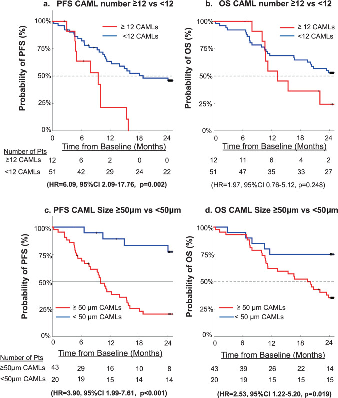
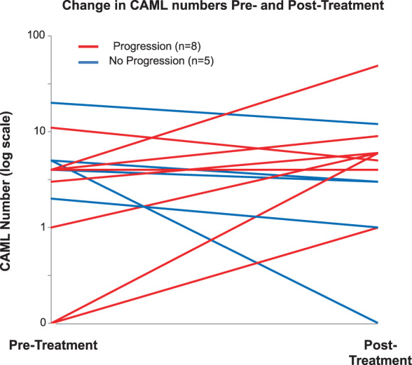
Pancreatic cancer (PC) is notoriously difficult to diagnosis and properly stage resulting in incorrect primary treatment. Diagnostic and prognostic biomarkers are desperately needed to more accurately stage patients and select proper treatments. Recently, a newly discovered circulating stromal cell, i.e. cancer associated macrophage-like cell (CAML), was found to accurately identify solid cancers and predict for worse prognosis. In this pilot study, blood samples were procured from 63 PC patients prior to start of therapeutic intent. CAMLs were found in 95% of samples tested, with ≥12 CAMLs/7.5 mL and ≥50 µm CAMLs both predicting for advanced pathological stage and progression free survival. These data suggest that CAML assessment prior to treatment of PC predicts patients with under-staged disease and with more aggressive PC less likely to respond to standard of care treatment.


Conflict of interest statement

The authors declare no competing non-financial interests but the following competing financial interests. D.L.A. and C.M.T. are employees of Creatv MicroTech, own stocks in Creatv MicroTech Inc. and have filed patent applications. K.P.G. is an independent contractor for Creatv MicroTech, Inc. The remaining authors have no other conflicts to report.

Figures

Fig. 1
Fig. 1. Images of CAML, CTC, and white blood cell.
ad CAMLs are enlarged cells with a polyploid nucleus (blue), diffuse Cytokeratin (green) and often CD45/CD14 positive (purple). eh CTCs are cells with nuclei(blue), filamented Cytokeratin (green), and are CD45/CD14 (purple) negative. White arrow highlights a normal white blood cell (DAPI and CD45/CD14 positive). Boxes = 75 µm.
Fig. 2
Fig. 2. Sensitivity and Specificity of CTCs and CAMLs in Pancreatic cancer patients referred for resection.
a Frequency of CTCs (>1 cell/sample), CEA (>5 ng/mL), CA19-9 (>37 µg/mL), and CAMLs (>1 cell/sample) as they relate to resectability. b ROC curve showing the specificity/sensitivity of CAMLs (red) and CTCs (blue) in comparison with 40 healthy controls.
Fig. 3
Fig. 3. CAML number and Largest CAML size based of Resectability and Pathological stage.
a Number of CAMLs found in patient baseline blood samples based on Pathological Stage and their original Resectability. Whisker Plots of CAML number based on Pathological Stage (median=red line). Wilcoxon ranked sum test of CAML number Stage 1 vs Stage 4 *p = 0.001, Stage 2 vs Stage 4 **p = 0.003, Stage 3 vs Stage 4 ***p = 0.015, and nonmetastatic vs metastatic disease ****p < 0.001. b Largest CAML found in patient baseline blood samples based on Pathological Stage and their original Resectability. Whisker Plots of CAML size based on Pathological Stage (median=red line). Wilcoxon ranked sum of CAML size Stage 1 vs Stage 4 *p < 0.001, Stage 2 vs Stage 4 **p = 0.010, Stage 3 vs Stage 4 ***p = 0.043 and nonmetastatic vs metastatic disease ****p < 0.001.
Fig. 4
Fig. 4. Kaplan–Meier graphs of PFS and OS for CAML number or size.
Using ≥ 12 CAMLs as a threshold, Kaplan Meier graphs for a PFS and b OS. Using ≥ 50 μm as a threshold for largest CAML size, Kaplan–Meier graphs for c PFS and d OS.
Fig. 5
Fig. 5. CAML enumeration for patients that progressed within 2 years versus patients that did not progress.
Patients who volunteered for multiple blood draws during the course of treatment (n = 13). Pre-Treatment and post-treatment time points were taken. Patients that experienced progression during a 2 year period (red lines), with six patients having an increase in CAML number, one patient having equal numbers, and one having a decrease in CAML number from 11 to 5 CAMLs. Patients that did not experience progression during a 2 year period (blue lines), with all five non-progressing patients had a decrease in CAML number.

References

    1. Howlader N. N. A. et al. SEER Cancer Statistics Review, 1975–2016, https://seer.cancer.gov/csr/1975_2016/ (2019).
    1. Ryan DP, Hong TS, Bardeesy N. Pancreatic adenocarcinoma. N. Engl. J. Med. 2014;371:2140–2141. doi: 10.1056/NEJMra1404198. - DOI - PubMed
    1. Reynolds RB, Folloder J. Clinical management of pancreatic cancer. J. Adv. Pr. Oncol. 2014;5:356–364. - PMC - PubMed
    1. Hartwig W, Werner J, Jager D, Debus J, Buchler MW. Improvement of surgical results for pancreatic cancer. Lancet Oncol. 2013;14:e476–e485. doi: 10.1016/S1470-2045(13)70172-4. - DOI - PubMed
    1. Stuart H, et al. Significant upstaging of patients with stage I pancreatic cancer from clinical to pathologic staging. J. Clin. Oncol. 2017;35:349–349. doi: 10.1200/JCO.2017.35.4_suppl.349. - DOI

LinkOut - more resources