Review
. 2021 May;124(10):1623-1636.
doi: 10.1038/s41416-021-01301-4. Epub 2021 Mar 19.

Pancreatic cancer cachexia: three dimensions of a complex syndrome

Affiliations
Review

Pancreatic cancer cachexia: three dimensions of a complex syndrome

Maximilian Kordes et al. Br J Cancer. 2021 May.

Abstract

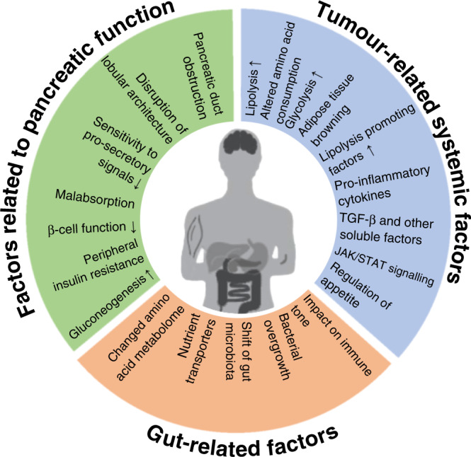
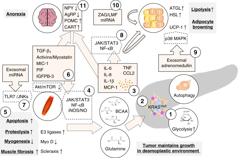
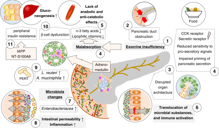
Cancer cachexia is a multifactorial syndrome that is characterised by a loss of skeletal muscle mass, is commonly associated with adipose tissue wasting and malaise, and responds poorly to therapeutic interventions. Although cachexia can affect patients who are severely ill with various malignant or non-malignant conditions, it is particularly common among patients with pancreatic cancer. Pancreatic cancer often leads to the development of cachexia through a combination of distinct factors, which, together, explain its high prevalence and clinical importance in this disease: systemic factors, including metabolic changes and pathogenic signals related to the tumour biology of pancreatic adenocarcinoma; factors resulting from the disruption of the digestive and endocrine functions of the pancreas; and factors related to the close anatomical and functional connection of the pancreas with the gut. In this review, we conceptualise the various insights into the mechanisms underlying pancreatic cancer cachexia according to these three dimensions to expose its particular complexity and the challenges that face clinicians in trying to devise therapeutic interventions.


Conflict of interest statement

M.K.: Alligator Bioscience (Consulting), Roche (Consulting); L.L.: no disclosures; L.E.: no disclosures; J.M.L.: Abbot (Honoraria), Mylan (Honoraria).

Figures

Fig. 1
Fig. 1. Conceptualisation of the three dimensions involved in the development of pancreatic cancer cachexia.
Pancreatic cancer cachexia is a complex syndrome that integrates three interdependent dimensions: tumour-related systemic factors; factors related to alterations of the pancreatic function; and factors related to the close interaction of the pancreas with other digestive organs. The systemic factors outlined are partly specific for cachexia associated with pancreatic adenocarcinoma, partly generic for that associated with solid malignancies. Factors related to the central function of the pancreas for nutritional uptake and homoeostasis, and factors related to other digestive organs are characteristic of pancreatic cancer cachexia.
Fig. 2
Fig. 2. Tumour-derived factors associated with cachexia in pancreatic adenocarcinoma.
Tumours increasingly metabolise glucose through glycolysis which is increased through mutant KRAS-dependent upregulation of glycolytic enzymes (1). Other KRAS-dependent metabolic changes promote the use of other carbon sources such as glutamine and branched chain amino acids (BCAA) from the breakdown of peripheral tissue, as well as other non-essential amino acids from pancreatic stellate cell autophagy, in the TCA cycle; substrates from the TCA cycle are used for biosynthesis (2). Together, these changes increase survival in a hypoxic and nutrient-deficient environment. Tumour-derived cytokines prompt several catabolic effects in peripheral tissues (3). Interleukin (IL)-6, TNF or interferon γ (IFNg) induce the breakdown of muscle fibres through the Janus kinase (JAK)/signal transducer and activator of transcription (STAT3) pathway and nuclear factor (NF)-κB-dependent induction of nitric oxide (4) which leads to decreased myogenesis through downregulation of the myogenic regulatory factor MyoD and increased proteolysis through E3-ligase-dependent ubiquitination (5). Similarly, members of the transforming growth factor (TGF)-β superfamily activate the canonical Smad2/3 pathway which promotes ubiquitin ligase-mediated proteolysis and scleraxis-mediated muscle fibrosis and reduces Akt/mTOR-mediated myogenesis (6). Additionally, microRNAs (miRNAs) in tumour-derived extracellular vesicles induce myoblast apoptosis by Toll-like receptor 7-dependent c-Jun N-terminal kinase (JNK) signalling (7). In white adipose tissue, pro-inflammatory signals, specifically IL-6, promote lipolysis through activation of the JAK/STAT3 pathway and NF-kB in adipose tissue and induces adipocyte browning through upregulation of uncoupling protein 1 (UCP-1) (8). Lipolysis through hormone-sensitive lipase (HSL) are also mediated by adrenomedullin from tumour-derived exosomes which activates the p38 mitogen-activated protein kinase (MAPK) pathway in adipocytes. Exosomal cargoes (other than adrenomedullin) can also induce adipocyte browning through UCP-1 expression (9). Lipolysis is maintained through paracrine signals in fat tissue liked zinc-α2-glycoprotein (ZAG/LMF) and miRNAs which induce adipose triglyceride lipase (ATGL) and HSL catalyse the hydrolysis of stored triglycerides (10). In the central nervous system, pro-inflammatory cytokines, particularly IL-1β, and macrophage inhibitory cytokine (MIC)-1 (a member of the TGF-β superfamily), induce anorexia by reducing anabolic neuropeptide Y (NPY)/agouti-related protein (AgRP) and increasing catabolic proopiomelanocortin (POMC) and cocaine-and-amphetamine regulated transcript (CART) signalling (11).
Fig. 3
Fig. 3. Impaired pancreatic exocrine and endocrine function interact with alterations in the digestive tract to promote pancreatic cancer cachexia.
Pancreatic cancer induces pancreatic exocrine insufficiency (PEI) (1) through obstruction of the pancreatic duct (2) or disruption of the organ architecture (3) as well as an altered physiological function, i.e. a reduced sensitivity to the pro-secretory signals secretin and cholecystokinin (CCK) resulting in an impaired priming of the pancreatic (4). PEI results in malabsorption of nutrients including lipophilic vitamins and n-3 fatty acids which results in insufficient caloric uptake and a lack of anabolic or anti-catabolic signals (5). PEI, in combination with changes of the histological architecture of the pancreas, also leads to excess bacterial growth in the duodenum and the translocation of bacteria and bacterial components into the tumour with implications for the immune tone (6). Pancreatic cancer is associated with specific changes of the gut microbiota and outgrowth of Enterobacteriaceae (7), particularly Klebsiella, is associated with increased intestinal permeability and systemic inflammation linked to the development of cachexia (8). Pancreatic enzyme replacement therapy (PERT) counteracts cancer-related dysbiosis and promotes the abundance of Lactobacillus reuteri and Akkermansia muciniphila which potentially reduce muscle wasting and increase the anti-tumoural immune response, respectively (9). Changes of the endocrine function of the pancreas comprise tumour-derived local adrenomedullin that reduces the Insulin secretion from islet cells in the pancreas (10) and an increase of peripheral insulin resistance through pancreatic-cancer-induced secretion of the islet amyloid polypeptide (IAPP) from β-cells and likely an increase of the S-100A8 N-terminal peptide (11). Endocrine dysregulation is also associated with increased gluconeogenesis (12), which depletes peripheral tissues to maintain the altered glucose utilisation.

References

    1. Fearon K, Strasser F, Anker SD, Bosaeus I, Bruera E, Fainsinger RL, et al. Definition and classification of cancer cachexia: an international consensus. Lancet Oncol. 2011;12:489–495. doi: 10.1016/S1470-2045(10)70218-7. - DOI - PubMed
    1. Baracos VE, Martin L, Korc M, Guttridge DC, Fearon KCH. Cancer-associated cachexia. Nat. Rev. Dis. Primers. 2018;4:17105. doi: 10.1038/nrdp.2017.105. - DOI - PubMed
    1. Martin L, Birdsell L, MacDonald N, Reiman T, Clandinin MT, McCargar LJ, et al. Cancer cachexia in the age of obesity: skeletal muscle depletion Is a powerful prognostic factor, independent of body mass index. J. Clin. Oncol. 2013;31:1539–1547. doi: 10.1200/JCO.2012.45.2722. - DOI - PubMed
    1. Minicozzi P, Cassetti T, Vener C, Sant M. Analysis of incidence, mortality and survival for pancreatic and biliary tract cancers across Europe, with assessment of influence of revised European age standardisation on estimates. Cancer Epidemiol. 2018;55:52–60. doi: 10.1016/j.canep.2018.04.011. - DOI - PubMed
    1. Hendifar AE, Chang JI, Huang BZ, Tuli R, Wu BU. Cachexia, and not obesity, prior to pancreatic cancer diagnosis worsens survival and is negated by chemotherapy. J. Gastrointest. Oncol. 2018;9:17–23. doi: 10.21037/jgo.2017.11.10. - DOI - PMC - PubMed

Publication types