

The mechanism of lncRNA-CRNDE in regulating tumour-associated macrophage M2 polarization and promoting tumour angiogenesis
- PMID: 33742511
- PMCID: PMC8093957
- DOI: 10.1111/jcmm.16477
The mechanism of lncRNA-CRNDE in regulating tumour-associated macrophage M2 polarization and promoting tumour angiogenesis
Erratum in
-
Corrigendum.J Cell Mol Med. 2022 Mar;26(5):1727-1728. doi: 10.1111/jcmm.17208. J Cell Mol Med. 2022. PMID: 35253381 Free PMC article. No abstract available.
Abstract
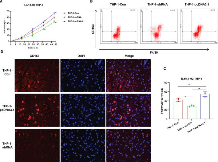
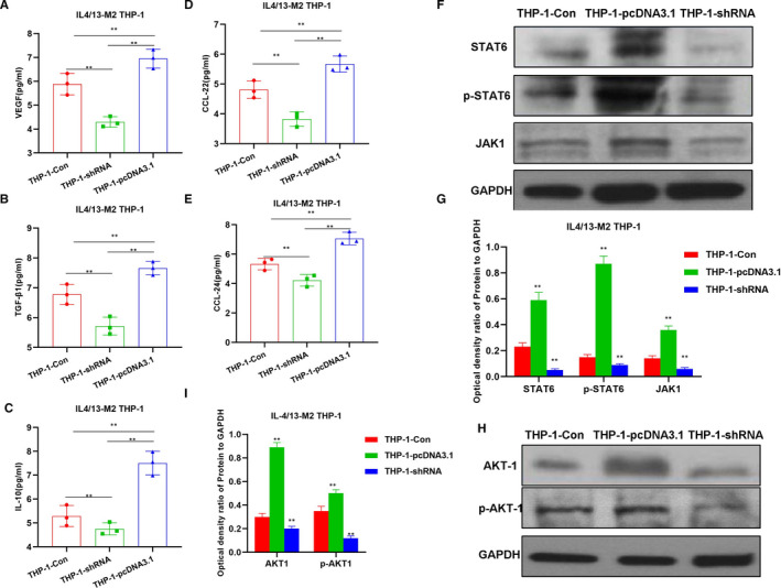
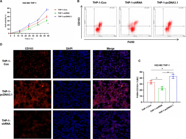
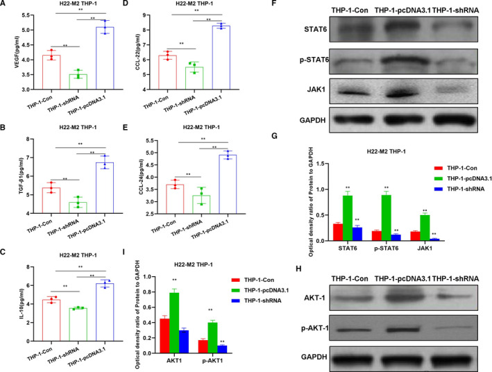
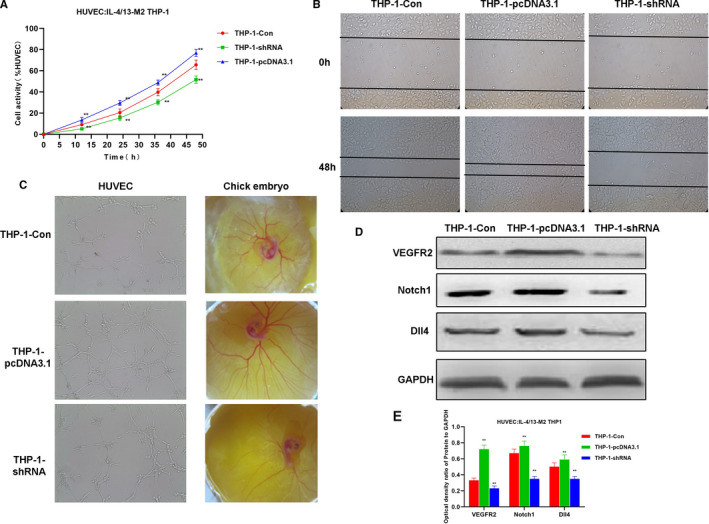
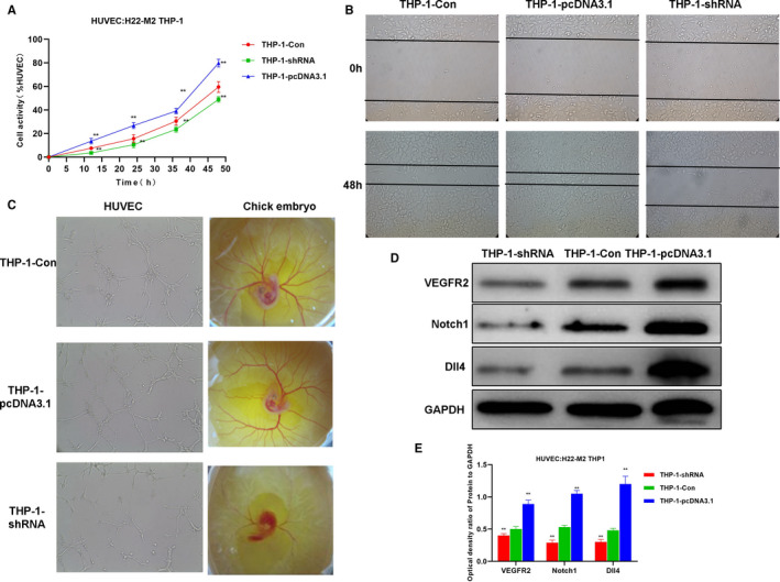
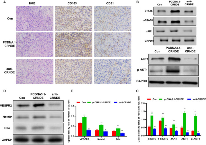
M2 macrophages can promote liver cancer metastasis by promoting tumour angiogenesis; however, the mechanism underlying macrophage polarization has not been completely revealed. In this study, we mainly explored the mechanism underlying long non-coding RNA-CRNDE (lncRNA-CRNDE) in regulating M2 macrophage polarization and promoting liver cancer angiogenesis. The expression of CRNDE was up-regulated or down-regulated in THP-1 cells (CRNDE-/- -THP-1 cells and pcDNA3.1-CRNDE-THP-1). THP-1 cells were co-cultured with liver cancer cell line H22, and M2 polarization was induced in THP-1 by IL-4/13 to simulate tumour-induced macrophage polarization. As a result, after CRNDE overexpression, THP-1 cell viability was up-regulated, the expression of M2 membrane marker CD163 was up-regulated, and the proportion of F4/80 + CD163+ cells was also up-regulated. ELISA assay showed that the expression of M2 markers (including TGF-β1 and IL-10) and chemokines (including CCl22 and CCL22) was up-regulated, and the expression of key signals (including STAT6, JAK-1, p-AKT1, and Arg-1) was also up-regulated, which were significantly different compared with the control group (Con). In addition, the intervention effect of CRNDE on THP-1 was consistent between co-culture with H22 cells and IL-4/13 induction assay. The induced M2 THP-1 cells were co-cultured with HUVEC. As a result, THP-1 cells with CRNDE overexpression can promote the migration and angiogenesis of HUVEC cells in vitro and simultaneously up-regulate the expression of Notch1, Dll4 and VEGFR2, indicating that THP-1 M2 polarization induced by CRNDE could further promote angiogenesis. The H22 cell tumour-bearing mouse model was constructed, followed by injection of CRNDE anti-oligosense nucleotides and overexpression plasmids to interfere CRNDE expression in tumour-bearing tissues. Consequently, down-regulation of CRNDE could down-regulate tumour volume, simultaneously down-regulate the expression of CD163 and CD31 in tissues, decrease the expression of key proteins (including JAK-1, STAT-6, p-STAT6 and p-AKT1), and down-regulate the expression of key angiogenesis-related proteins (including VEGF, Notch1, Dll4 and VEGFR2). In this study, we found that CENDE could indirectly regulate tumour angiogenesis by promoting M2 polarization of macrophages, which is also one of the mechanisms of microenvironmental immune regulation in liver cancer.
Keywords: M2 polarization; long non-coding RNA-CRNDE; macrophages; microenvironment; tumour blood vessels.
© 2021 The Authors. Journal of Cellular and Molecular Medicine published by Foundation for Cellular and Molecular Medicine and John Wiley & Sons Ltd.
Conflict of interest statement
No competing interests.
Figures
Similar articles
-
Long non-coding RNA GAS5 overexpression inhibits M2-like polarization of tumour-associated macrophages in SMCC-7721 cells by promoting PTEN expression.Int J Exp Pathol. 2020 Dec;101(6):215-222. doi: 10.1111/iep.12374. Epub 2020 Nov 4. Int J Exp Pathol. 2020. PMID: 33146930 Free PMC article.
-
lncRNA CRNDE promotes the proliferation and metastasis by acting as sponge miR-539-5p to regulate POU2F1 expression in HCC.BMC Cancer. 2020 Apr 6;20(1):282. doi: 10.1186/s12885-020-06771-y. BMC Cancer. 2020. PMID: 32252678 Free PMC article.
-
Long Non-Coding RNA CRNDE Regulates Angiogenesis in Hepatoblastoma by Targeting the MiR-203/VEGFA Axis.Pathobiology. 2020;87(3):161-170. doi: 10.1159/000505131. Epub 2020 Mar 17. Pathobiology. 2020. PMID: 32182608
-
Macrophage polarization in hepatocellular carcinoma: a lncRNA-centric perspective on tumor progression and metastasis.Clin Exp Med. 2025 May 25;25(1):173. doi: 10.1007/s10238-025-01711-1. Clin Exp Med. 2025. PMID: 40413657 Free PMC article. Review.
-
Long Non-coding RNAs Regulating Macrophage Polarization in Liver Cancer.Curr Pharm Des. 2024;30(27):2120-2128. doi: 10.2174/0113816128311861240523075218. Curr Pharm Des. 2024. PMID: 38859791 Review.
Cited by
-
Integrated Analysis of Multi-Omics Data to Establish a Hypoxia-Related Prognostic Model in Osteosarcoma.Evol Bioinform Online. 2022 Oct 26;18:11769343221128537. doi: 10.1177/11769343221128537. eCollection 2022. Evol Bioinform Online. 2022. PMID: 36325183 Free PMC article.
-
New Angiogenic Regulators Produced by TAMs: Perspective for Targeting Tumor Angiogenesis.Cancers (Basel). 2021 Jun 29;13(13):3253. doi: 10.3390/cancers13133253. Cancers (Basel). 2021. PMID: 34209679 Free PMC article. Review.
-
Crosstalk among long non-coding RNA, tumor-associated macrophages and small extracellular vesicles in tumorigenesis and dissemination.Front Oncol. 2022 Oct 3;12:1008856. doi: 10.3389/fonc.2022.1008856. eCollection 2022. Front Oncol. 2022. PMID: 36263199 Free PMC article. Review.
-
Amino acid metabolism regulated by lncRNAs: the propellant behind cancer metabolic reprogramming.Cell Commun Signal. 2023 May 1;21(1):87. doi: 10.1186/s12964-023-01116-1. Cell Commun Signal. 2023. PMID: 37127605 Free PMC article. Review.
-
LncRNAs in oncogenic microenvironment: from threat to therapy.Front Cell Dev Biol. 2025 Mar 13;12:1423279. doi: 10.3389/fcell.2024.1423279. eCollection 2024. Front Cell Dev Biol. 2025. PMID: 40176927 Free PMC article. Review.
References
-
- Mantovani A, Sozzani S, Locati M, et al. Macrophage polarization: tumor‐associated macrophages as a paradigm for polarized M2 mononuclear phagocytes. Trends Immunol. 2002;23(11):549‐555. - PubMed
-
- Relation T, Yi BT, Guess CAJ, et al. Intratumoral delivery of interferon‐secreting mesenchymal stromal cells repolarizes tumor‐associated macrophages and suppresses neuroblastoma proliferation in vivo. Stem Cells. 2018;36(6):915‐924. - PubMed
-
- Han SC, Koo DH, Kang NJ, et al. The generation of Tregs and IL‐10/TGF‐β‐modified macrophages by docosahexaenoic acid via TGF‐β dependent mechanism suppresses atopic dermatitis. Cytokine. 2014;70(1):44.
Publication types
MeSH terms
Substances
Grants and funding
LinkOut - more resources
Full Text Sources
Other Literature Sources
Medical
Research Materials
Miscellaneous
