. 2021 Jul;14(4):1490-1504.
doi: 10.1111/cts.13012. Epub 2021 May 31.

Identification of small molecules that mitigate vincristine-induced neurotoxicity while sensitizing leukemia cells to vincristine

Affiliations

Identification of small molecules that mitigate vincristine-induced neurotoxicity while sensitizing leukemia cells to vincristine

Barthelemy Diouf et al. Clin Transl Sci. 2021 Jul.

Abstract

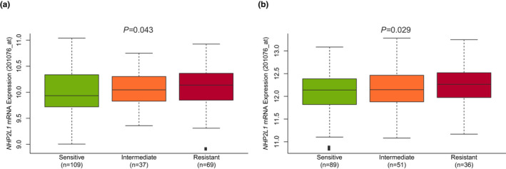
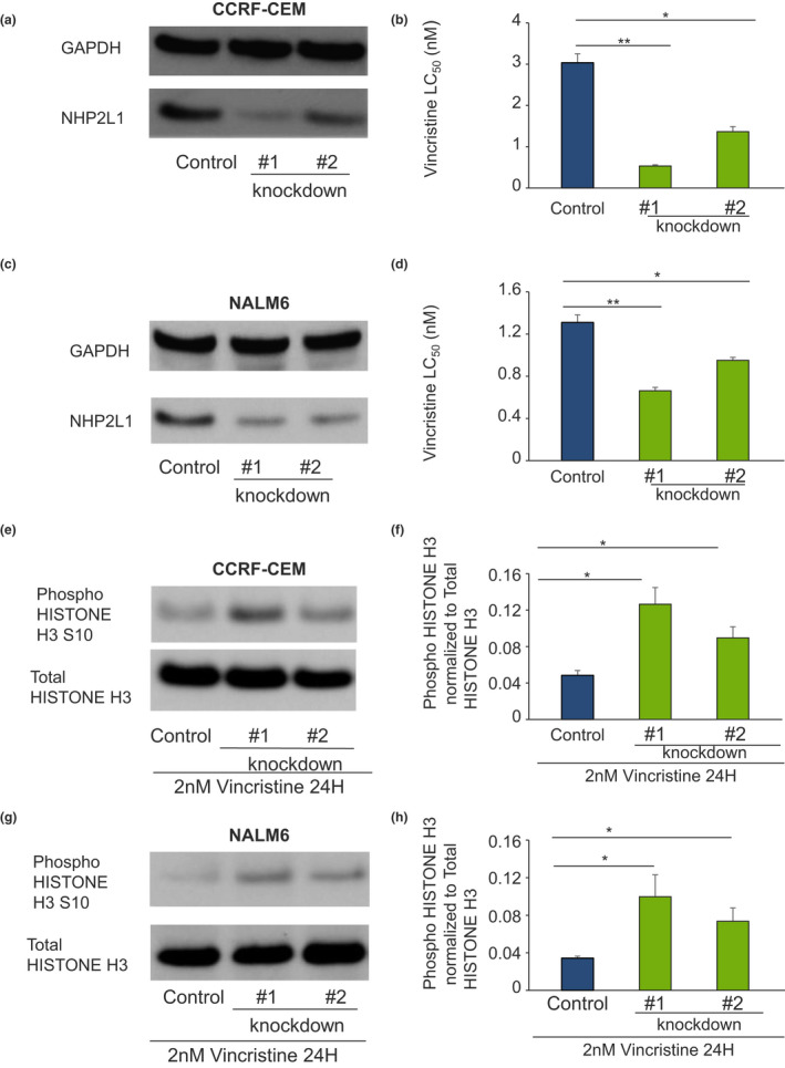
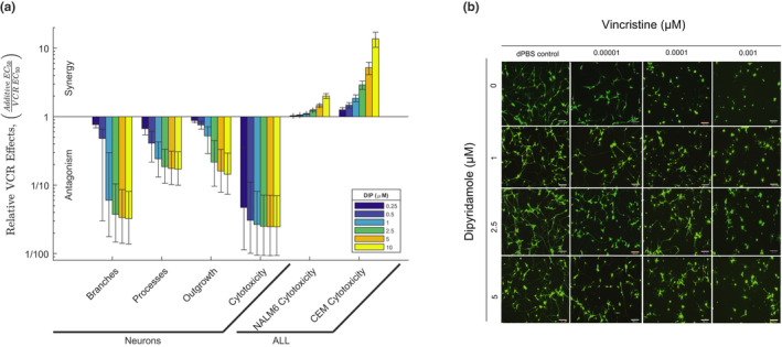
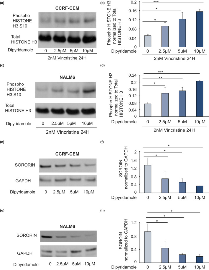
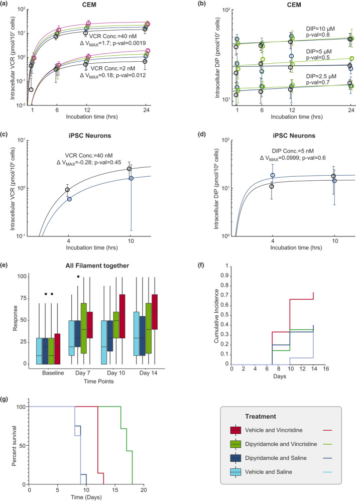
Vincristine (VCR) is one of the most widely prescribed medications for treating solid tumors and acute lymphoblastic leukemia (ALL) in children and adults. However, its major dose-limiting toxicity is peripheral neuropathy that can disrupt curative therapy. Peripheral neuropathy can also persist into adulthood, compromising quality of life of childhood cancer survivors. Reducing VCR-induced neurotoxicity without compromising its anticancer effects would be ideal. Here, we show that low expression of NHP2L1 is associated with increased sensitivity of primary leukemia cells to VCR, and that concomitant administration of VCR with inhibitors of NHP2L1 increases VCR cytotoxicity in leukemia cells, prolongs survival of ALL xenograft mice, but decreases VCR effects on human-induced pluripotent stem cell-derived neurons and mitigates neurotoxicity in mice. These findings offer a strategy for increasing VCR's antileukemic effects while reducing peripheral neuropathy in patients treated with this widely prescribed medication.


Conflict of interest statement

The authors declared no competing interests for this work.

Figures

FIGURE 1
FIGURE 1
NHP2L1 expression and sensitivity of primary B leukemic cells to vincristine (VCR). NHP2L1 mRNA expression was determined by Affymetrix gene expression array. MTT was used to determine the sensitivity of primary cells to VCR. The association between log2 expression of NHP2L1 and VCR sensitivity was assessed in two cohorts of patients with acute lymphoblastic leukemia: (a) St. Jude Children’s Research Hospital and (b) Dutch Childhood Oncology Group (DCOG)/Cooperative Study Group for Childhood Acute Lymphoblastic Leukemia (COALL) cohorts. Based on previously published criteria24 sensitive cells were defined as having a VCR concentration lethal to 50% of the cells (LC50) of 0.391 μg/ml (0.474 µM) or less, and resistant cells were defined as having an LC50 of 1.758 μg/ml (2.131 µM) or more. The horizontal line inside each box depicts the median, the upper and lower limits of the box are the 75th and 25th percentiles, respectively, and the vertical bars above and below each box indicate the maximum and minimum values, respectively. The p values were determined by linear regression comparing NHP2L1 gene expression and leukemia cell sensitivity to VCR
FIGURE 2
FIGURE 2
Knockdown of NHP2L1 and sensitivity to vincristine (VCR) and mitotic arrest in leukemic cell lines. Human leukemia cells infected with two different shRNAs against NHP2L1 show a decrease in NHP2L1 protein levels in CCRF‐CEM (a) and in NALM6 (c). After knockdown of NHP2L1, cells were treated for 72 h in the presence of increasing concentrations of VCR. VCR sensitivity (lethal concentration 50% [LC50]) Cell viability determined by MTT in CCRF‐CEM (b) and in NALM6 (d) are represented. Error bars represent SDs of three replicate experiments. Student’s t‐test was used to calculate p values. *p < 0.05, **p < 0.01. Human leukemia cells (CCRF‐CEM and NALM6) were infected with two different shRNAs (#1 and #2) against NHP2L1 and treated with vincristine for 24 h. Representative blots of phosphorylated Serine 10 in Histone H3 protein and of total Histone H3 after knockdown of NHP2L1 and 24‐h treatment with VCR were shown in CEM (e) and NALM6 cells (g). Phospho Histone H3 protein levels were quantified by densitometry, normalized to total Histone H3 signal and expressed in arbitrary units for the CEM cells (f) and for the NALM6 cells (h). Values are means ± SD of three independent experiments. Student’s t‐test was used to calculate p values. *p < 0.05, **p < 0.01
FIGURE 3
FIGURE 3
Effects of small molecule inhibitor of NHP2L1 U4 interaction on neurons and leukemic cells treated with vincristine (VCR). (a) VCR effects relative to dipyridamole (DIP) on neurons and two acute lymphoblastic leukemia (ALL) cell lines (CEM and NALM6). The y‐axis represents the ratio of the additive half‐maximal effective concentration (EC50) to the actual EC50 for the combination of VCR and DIP. Each bar represents a fixed concentration of DIP (0.25 µM [0.125 mg/L], 0.5 µM [0.25 mg/L], 1 µM [0.5 mg/L], 2.5 µM [1.265 mg/L], 5 µM [2.525 mg/L], and 10 µM [5.05 mg/L]). The whiskers are the 95% confidence interval of the measure. A ratio of one indicates the combination is additive, whereas a ratio greater or less than one indicates that the combination is either synergistic or antagonistic, respectively. (b) Representative images of human‐induced pluripotent stem cell (hiPSC) neurons (Peri.4 U) 72 h after treatment with increasing concentrations of VCR with or without increasing concentrations of DIP. Images were taken from the ImageXpress Micro imaging microscope at 10× magnification
FIGURE 4
FIGURE 4
Effects of dipyridamole (DIP) on vincristine (VCR)‐induced mitotic arrest and on SORORIN protein level. Human leukemia cells (CCRF‐CEM and NALM6) were treated with vincristine (2 nM [1.65 µg/L]) for 24 h alone or with increasing concentrations of DIP. Representative blots of phosphorylated Serine 10 in Histone H3 protein and of total Histone H3 after 24‐h treatment with VCR and increasing concentrations of DIP for human acute lymphoblastic leukemia cell lines CEM (a) and NALM6 (c) are shown. Phospho Serine 10 Histone H3 protein levels were quantified by densitometry, normalized to total Histone H3 signal and expressed in arbitrary units for the CEM cells (b) and for the NALM6 cells (d). Values are means ± SD of three independent experiments. Student’s t‐test was used to calculate p values. *p < 0.05, **p < 0.01, ***p < 0.001. G2 synchronized cells were treated with increasing concentration of DIP, and expression of SORORIN was document by Western blot in CEM (e) and NALM6 (g) cells. The level of SORORIN was normalized to the level of GAPDH and the ratio was plotted for the CEM (f) and the NALM6 (h) cells. Values are means ±SD of three independent experiments. Student’s t‐test was used to calculate p values. *p < 0.05, **p < 0.01, ***p < 0.001
FIGURE 5
FIGURE 5
Intracellular concentration of vincristine (VCR) or dipyridamole (DIP) in CEM human leukemia cell line, or human‐induced pluripotent stem cell (hiPSC)‐derived neurons relative to incubation time (hours) and VCR neurotoxicity in mice. Intracellular concentration of VCR was determined in (a) CEM acute lymphoblastic leukemia (ALL) cells after exposure of the cells to 2 nM (1.65 µg/L) or 40 nM (33 µg/L) of VCR with or without increasing concentrations of DIP: 0 µM in black, 2.5 µM (1.265 mg/L) in blue, 5 µM (2.525 mg/L) in green, and 10 µM (5.05 mg/L) in magenta. The circles and whiskers represent the median and range of the data. The maximal rate of metabolism (ΔVmax ) represents the change in Vmax due to the presence of DIP. The p values test whether the change in Vmax (ΔVmax ) due to the presence of DIP differs from 0 using the t‐test (a). Intracellular concentration of DIP was determined in (b) CEM after treating leukemia cells with 2.5 µM (1.265 mg/L), 5 µM (2.525 mg/L), or 10 µM (5.05 mg/L) of DIP with or without increasing concentrations of VCR: 0 nM in black, 2 nM (1.65 µg/L) in blue, and 40 nM (33 µg/L) in green. The circles and whiskers represent the median and range of the data. The p values test whether the change in the slope due to the presence of VCR is different from 0 using the t‐test (b). Intracellular levels of DIP were not significantly related to extracellular VCR concentrations. Intracellular concentration of VCR was determined in human‐induced pluripotent stem cell (hiPSC)‐derived neurons after treatment of the cells with 40 nM (33 µg/L) of VCR with or without 5 µM (2.525 mg/L) of DIP: 0 µM in black, and 5 µM (2.525 mg/L) in blue (c). The p values test if ∆Vmax differs due to the presence or absence of dipyridamole using the t‐test (c). The circles and whiskers represent the median and range of the data. The ΔVmax represents the change in Vmax due to the presence of DIP. Intracellular concentration of DIP was determined in hiPSC‐derived neurons after treating the cells with 5 µM of DIP with or without 40 nM (33 µg/L) of VCR: 0 nM in black, and 40 nM (33 µg/L) in blue. The circles and whiskers represent the median and range of the data (d). The p values test whether ∆Vmax due to the presence of vincristine is different from 0 using the t‐test (d). (e) Paw withdrawal responses expressed as percentage were determined using von Frey filaments at baseline, day 7, day 10 and day 14 in mice receiving one of the following treatments: saline and vehicle of DIP; saline and DIP; VCR and vehicle of DIP; and VCR and DIP. (f) Cross‐sectional and cumulative proportion (CPN) of mice positive for neurotoxicity in each treatment group is depicted. Positive neurotoxicity was defined as values higher than 95th percentile of the von Frey measurements. (g) NSG mice were inoculated with human CEM leukemia cells. After engraftment, the mice were treated once a week with saline and vehicle (controls), VCR, DIP, or the two drugs together. Percentage survival was determined for each group of mice. Mice receiving VCR and vehicle showed a significantly prolonged survival compared with controls (saline + vehicle as well as saline + dipyridamole; p value <0.001). The addition of DIP to VCR significantly prolonged the survival of mice compared with mice treated with only VCR and vehicle (p value <0.001)

Similar articles

Cited by

References

    1. Calcabrini C, Maffei F, Turrini E, Fimognari C. Sulforaphane potentiates anticancer effects of doxorubicin and cisplatin and mitigates their toxic effects. Front Pharmacol. 2020;11:567. - PMC - PubMed
    1. Alam M, Yadav RK, Minj E, Tiwari A, Mehan S. Exploring molecular approaches in Amyotrophic lateral sclerosis: drug targets from clinical and pre‐clinical findings [published online ahead of print April 27, 2020). Curr Molec Pharmacol. https://doi.org/10.2174/1566524020666200427214356. - DOI - PubMed
    1. Bradley WG, Lassman LP, Pearce GW, Walton JN. The neuromyopathy of vincristine in man. Clinical, electrophysiological and pathological studies. J Neurol Sci. 1970;10:107‐131. - PubMed
    1. Gidding CEM, Meeuwsen‐de Boer GJ, Koopmans P, Uges DRA, Kamps WA, de Graaf SSN. Vincristine pharmacokinetics after repetitive dosing in children. Cancer Chemother. Pharmacol. 1999;44:203‐209. - PubMed
    1. Bjornard KL, Gilchrist LS, Inaba H, et al. Peripheral neuropathy in children and adolescents treated for cancer. Lancet Child Adolesc Health. 2018;2:744‐754. - PMC - PubMed

Publication types

MeSH terms

LinkOut - more resources