

Lung ultrasound for the early diagnosis of COVID-19 pneumonia: an international multicenter study
- PMID: 33743018
- PMCID: PMC7980130
- DOI: 10.1007/s00134-021-06373-7
Lung ultrasound for the early diagnosis of COVID-19 pneumonia: an international multicenter study
Abstract
Purpose: To analyze the application of a lung ultrasound (LUS)-based diagnostic approach to patients suspected of COVID-19, combining the LUS likelihood of COVID-19 pneumonia with patient's symptoms and clinical history.
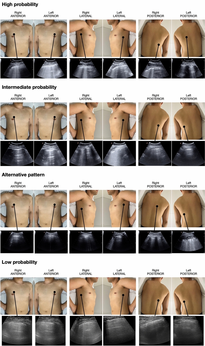
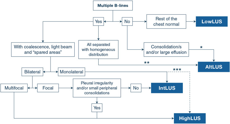
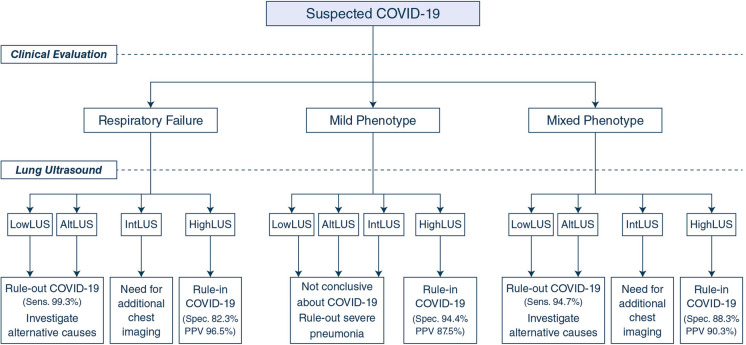
Methods: This is an international multicenter observational study in 20 US and European hospitals. Patients suspected of COVID-19 were tested with reverse transcription-polymerase chain reaction (RT-PCR) swab test and had an LUS examination. We identified three clinical phenotypes based on pre-existing chronic diseases (mixed phenotype), and on the presence (severe phenotype) or absence (mild phenotype) of signs and/or symptoms of respiratory failure at presentation. We defined the LUS likelihood of COVID-19 pneumonia according to four different patterns: high (HighLUS), intermediate (IntLUS), alternative (AltLUS), and low (LowLUS) probability. The combination of patterns and phenotypes with RT-PCR results was described and analyzed.
Results: We studied 1462 patients, classified in mild (n = 400), severe (n = 727), and mixed (n = 335) phenotypes. HighLUS and IntLUS showed an overall sensitivity of 90.2% (95% CI 88.23-91.97%) in identifying patients with positive RT-PCR, with higher values in the mixed (94.7%) and severe phenotype (97.1%), and even higher in those patients with objective respiratory failure (99.3%). The HighLUS showed a specificity of 88.8% (CI 85.55-91.65%) that was higher in the mild phenotype (94.4%; CI 90.0-97.0%). At multivariate analysis, the HighLUS was a strong independent predictor of RT-PCR positivity (odds ratio 4.2, confidence interval 2.6-6.7, p < 0.0001).
Conclusion: Combining LUS patterns of probability with clinical phenotypes at presentation can rapidly identify those patients with or without COVID-19 pneumonia at bedside. This approach could support and expedite patients' management during a pandemic surge.
Keywords: COVID-19; Interstitial pneumonia; Lung ultrasound; SARS-CoV-2.
Conflict of interest statement
All the authors have had access to all the data of the study and accept responsibility for its validity. None of the authors and collaborators has conflicts of interest related to the present paper to diclose.
Figures
Comment in
-
Limiting the areas inspected by lung ultrasound leads to an underestimation of COVID-19 patients' condition.Intensive Care Med. 2021 Jul;47(7):811-812. doi: 10.1007/s00134-021-06407-0. Epub 2021 May 11. Intensive Care Med. 2021. PMID: 33974109 Free PMC article. No abstract available.
-
Determining the optimal number of lung ultrasound zones to monitor COVID-19 patients: can we keep it ultra-short and ultra-simple?Intensive Care Med. 2021 Sep;47(9):1041-1043. doi: 10.1007/s00134-021-06463-6. Epub 2021 Jun 26. Intensive Care Med. 2021. PMID: 34173859 Free PMC article. No abstract available.
References
-
- Lichter Y, Topilsky Y, Taieb P, Banai A, Hochstadt A, Merdler I, Gal Oz A, Vine J, Goren O, Cohen B, Sapir O, Granot Y, Mann T, Friedman S, Angel Y, Adi N, Laufer-Perl M, Ingbir M, Arbel Y, Matot I, Szekely Y. Lung ultrasound predicts clinical course and outcomes in COVID-19 patients. Intensive Care Med. 2020;46:1873–1883. doi: 10.1007/s00134-020-06212-1. - DOI - PMC - PubMed
-
- de Almeida Monteiro RA, Duarte-Neto AN, Ferraz da Silva LF, de Oliveira EP, do Nascimento ECT, Mauad T, Saldiva PHDN, Dolhnikoff M. Ultrasound assessment of pulmonary fibroproliferative changes in severe COVID-19: a quantitative correlation study with histopathological findings. Intensive Care Med. 2021;3:1–9. doi: 10.1007/s00134-020-06328-4. - DOI - PMC - PubMed
-
- Zieleskiewicz L, Markarian T, Lopez A, Taguet C, Mohammedi N, Boucekine M, Baumstarck K, Besch G, Mathon G, Duclos G, Bouvet L, Michelet P, Allaouchiche B, Chaumoître K, Di Bisceglie M, Leone M, Network AZUREA. Comparative study of lung ultrasound and chest computed tomography scan in the assessment of severity of confirmed COVID-19 pneumonia. Intensive Care Med. 2020;46:1707–1713. doi: 10.1007/s00134-020-06186-0. - DOI - PMC - PubMed
-
- Volpicelli G, Elbarbary M, Blaivas M, Lichtenstein DA, Mathis G, Kirkpatrick AW, Melniker L, Gargani L, Noble VE, Via G, Dean A, Tsung JW, Soldati G, Copetti R, Bouhemad B, Reissig A, Agricola E, Rouby JJ, Arbelot C, Liteplo A, Sargsyan A, Silva F, Hoppmann R, Breitkreutz R, Seibel A, Neri L, Storti E, Petrovic T, International Liaison Committee on Lung Ultrasound (ILC-LUS) for International Consensus Conference on Lung Ultrasound (ICC-LUS) International evidence-based recommendations for point-of-care lung ultrasound. Intensive Care Med. 2012;38:577–591. doi: 10.1007/s00134-012-2513-4. - DOI - PubMed
-
- Laursen CB, Sloth E, Lassen AT, Christensen Rd, Lambrechtsen J, Madsen PH, Henriksen DP, Davidsen JR, Rasmussen F. Point-of-care ultrasonography in patients admitted with respiratory symptoms: a single-blind, randomised controlled trial. Lancet Respir Med. 2014;2:638–646. doi: 10.1016/S2213-2600(14)70135-3. - DOI - PubMed
Publication types
MeSH terms
LinkOut - more resources
Full Text Sources
Other Literature Sources
Medical
Miscellaneous
