. 2021 Apr 29;184(9):2372-2383.e9.
doi: 10.1016/j.cell.2021.03.013. Epub 2021 Mar 12.

Multiple SARS-CoV-2 variants escape neutralization by vaccine-induced humoral immunity

Affiliations

Multiple SARS-CoV-2 variants escape neutralization by vaccine-induced humoral immunity

Wilfredo F Garcia-Beltran et al. Cell. .

Erratum in

Abstract

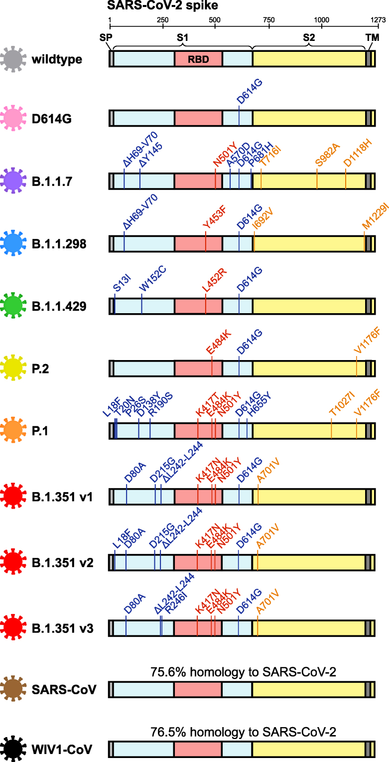
Vaccination elicits immune responses capable of potently neutralizing SARS-CoV-2. However, ongoing surveillance has revealed the emergence of variants harboring mutations in spike, the main target of neutralizing antibodies. To understand the impact of these variants, we evaluated the neutralization potency of 99 individuals that received one or two doses of either BNT162b2 or mRNA-1273 vaccines against pseudoviruses representing 10 globally circulating strains of SARS-CoV-2. Five of the 10 pseudoviruses, harboring receptor-binding domain mutations, including K417N/T, E484K, and N501Y, were highly resistant to neutralization. Cross-neutralization of B.1.351 variants was comparable to SARS-CoV and bat-derived WIV1-CoV, suggesting that a relatively small number of mutations can mediate potent escape from vaccine responses. While the clinical impact of neutralization resistance remains uncertain, these results highlight the potential for variants to escape from neutralizing humoral immunity and emphasize the need to develop broadly protective interventions against the evolving pandemic.

Keywords: COVID-19; RBD; SARS-CoV-2; escape; mRNA vaccines; neutralizing antibodies; spike; variants.


Conflict of interest statement

Declaration of interests The authors declare no competing interests.

Figures

None
Graphical abstract
Figure 1
Figure 1
Emergence and spread of SARS-CoV-2 variants of concern around the world (A) Phylogenetic tree of SARS-CoV-2 variants (adapted from nextstrain.org) with sampling dates is illustrated with a focus on the following lineages: A (gray), B.1.1.7 (purple), B.1.1.298 (blue), B.1.429 (green), P.2 (yellow), P.1 (orange), and B.1.351 (red). Dotted lines to SARS-CoV (brown) and bat-derived WIV1-CoV (black) are not to scale but indicate a distant phylogenetic relationship to SARS-CoV-2. (B) World map depicting the locations where the variants of these lineages were first described: original wild-type virus from A lineage (gray) in Wuhan, China; D614G variant (pink) in Europe that became dominant circulating strain; B.1.1.7 lineage (purple) in the United Kingdom; B.1.1.298 (blue) in Denmark; B.1.429 (green) in California, United States; P.2 (yellow) in Brazil and Japan; P.1 (orange) in Brazil and Japan; and B.1.351 (red) in South Africa. (C) Crystal structure of pre-fusion stabilized SARS-Cov-2 spike trimer (PDB: 7JJI) is shown with top (left panel) and side (right panel) views. Sites where naturally occurring mutations occur are indicated with residue atoms highlighted as colored spheres. Spike regions and associated mutations are colored as follows: receptor binding domain (RBD) in red, S1 (excluding RBD) in blue, and S2 in yellow. (D) Schematic of SARS-CoV-2 spike protein structure and the mutation landscape of variants used in this study are illustrated. The mutations present in each variant tested represent the consensus sequence for that lineage and represent actual circulating strains: A (wild type), B.1.1.7, B.1.1.298, B.1.1.429, P.2, and P.1 lineages. In the case of the B.1.351 lineage, the three most abundant variants (v1, v2, and v3) deposited in GISAID were assessed. For SARS-CoV and WIV1-CoV, the percent homology is indicated. The following abbreviations are used: SP, signal peptide; TM, transmembrane domain; RBD, receptor binding domain. In the mutation map, a dot () indicates the same amino acid in that position as wild type and a dash () indicates a deletion.
Figure S1
Figure S1
SARS-CoV-2 variants tested in this study, related to Figure 1 Schematic of mutations in the spike protein sequence of the following SARS-CoV-2 variants are illustrated: wild type (gray), D614G (pink), B.1.1.7 (purple), B.1.1.298 (blue), B.1.1.429 (green), P.2 (yellow), P.1 (orange), three variants of B.1.351 (red; v1, v2, and v3), SARS-CoV (brown), and WIV1-CoV (black).
Figure 2
Figure 2
Neutralization of SARS-CoV-2 pseudovirus variants by vaccinee sera (A) Schematic of our vaccine recipient cohort, consisting of individuals who received one or two full doses of either BNT162b2 (Pfizer) or mRNA-1273 (Moderna) vaccine, is presented in conjunction with our previously described high-throughput lentiviral vector-based SARS-CoV-2 pseudovirus neutralization assay (Garcia-Beltran et al., 2021). (B) Representative pseudovirus neutralization curves are shown for an individual ≥7 days out from the second dose of BNT162b2 vaccine comparing wild-type SARS-CoV-2 pseudovirus to the following variant pseudoviruses: D614G (pink); B.1.1.7 (purple); B.1.1.298 (blue); B.1.429 (green); P.2 (yellow); P.1 (orange); B.1.351 v1, v2, and v3 (red); SARS-CoV (brown); and WIV1-CoV (black). Dots and error bars indicate mean and standard deviation.
Figure S2
Figure S2
Characteristics of vaccine recipients, related to Figure 2 The sex and age of 99 vaccine recipients are presented along with time (in days) after the first and, when applicable, second doses of either the BNT162b2 (left panel) or mRNA-1273 (right panel) vaccines. Individuals who reported having been diagnosed with or highly suspected to have COVID-19 before vaccination are highlighted with a red circle. For 7 out of the 99 vaccinees, two samples taken, one after the first dose and one after the second dose.
Figure S3
Figure S3
Receiver operating characteristic analysis of pseudovirus neutralization assay in vaccinated and non-vaccinated individuals, related to Figure 3 (A and B) Receiver operating curve (ROC) analysis was performed in non-vaccinated pre-pandemic individuals (n = 1,220) and recipients of 2 full doses of either BNT162b2 or mRNA-1273 (2-dose vaccinated, n = 65) (A) or 1 dose (or < 7 days from second dose) of either vaccine (1-dose vaccinated, n = 41) (B). Area under the curve (AUC) is presented.
Figure 3
Figure 3
COVID-19 vaccines elicit potent but dose-dependent neutralizing responses against SARS-CoV-2 regardless of age or sex (A) Titers that achieve 50% pseudovirus neutralization (pNT50) of wild-type SARS-CoV-2 are plotted for the following cohorts: (1) pre-pandemic individuals who were never vaccinated or infected with SARS-CoV-2 (pre-pandemic, white circles, n = 1,220), (2) vaccine recipients that received only one dose of the BNT162b2 vaccine or were <7 days from their second dose (BNT162b2 – one dose, light blue triangles, n = 14), (3) vaccine recipients ≥7 days following their second dose of BNT162b2 vaccine (BNT162b2 – two dose, blue circles, n = 30), (4) vaccine recipients that received only one dose of the mRNA-1273 vaccine or were <7 days from their second dose (mRNA-1273 – one dose, pink diamonds, n = 27), and (5) vaccine recipients ≥7 days following their second dose of the mRNA-1273 vaccine (mRNA-1273 – two dose, red squares, n = 35). (B) Pseudovirus neutralization (pNT50) of wild-type SARS-CoV-2 is plotted versus time post-vaccination. The first dose (“dose #1”) of both BNT162b2 (blue) and mRNA-1273 (red) vaccines occurred on day 0. The second dose of the BNT162b2 vaccine occurred approximately 21 days after the first dose (“dose #2” in blue text) and for the mRNA-1273 vaccine occurred approximately 29 days after the first dose (“dose #2” in red text). Groups of vaccinees are the same as in (A). (C) Total anti-RBD (wild type) antibody levels measured by a quantitative ELISA against the wild-type RBD antigen versus neutralization of wild-type SARS-CoV-2 pseudovirus are presented. ELISAs were performed in duplicate and average values were used. Groups of vaccinees are the same as in (A). (D) Neutralization of wild-type SARS-CoV-2 pseudovirus is compared between female and male vaccinees. Bars and error bars indicate mean and standard deviation. Groups of vaccinees are the same as in (A). (E) Neutralization of wild-type SARS-CoV-2 pseudovirus is plotted against age of vaccine recipient. Groups of vaccinees are the same as in (A).
Figure 4
Figure 4
Sera from COVID-19 vaccine recipients cross-neutralize some but not all SARS-CoV-2 variants of concern (A and B) Pseudovirus neutralization (pNT50) is plotted for all individuals that received one dose (bottom panels) or two full doses (upper panels) of either the BNT162b2 (A) or mRNA-1273 (B) vaccines for each of the following SARS-CoV-2 pseudoviruses: wild-type, D614G, B.1.1.7, B.1.1.298, B.1.429, P.2, P.1, and three variants of the B.1.351 lineage denoted as B.1.351 v1, v2, and v3. Other distantly related coronaviruses, namely, SARS-CoV from the 2002 Hong Kong outbreak and the pre-emergent bat coronavirus WIV1-CoV, were also tested. Individuals who were <7 days out from their second dose of the vaccine were classified as having received one dose. Dotted lines indicate vaccine recipients that were previously diagnosed with or highly suspected to have COVID-19 before being vaccinated. Gray regions indicate upper and lower limits of detection of our neutralization assay. The following abbreviations are used to indicate the country where the variant was first described: UK, United Kingdom; DK, Denmark; US, United States; BR, Brazil; JP, Japan; and SA, South Africa. An ANOVA correcting for multiple comparisons was performed, and statistical significance of each pseudovirus relative to wild type is shown with the following notations: p < 0.05, ∗∗∗p < 0.001, ∗∗∗∗p < 0.0001. (C and D) Fold decrease in neutralization for each pseudovirus relative to wild type is shown for vaccine recipients ≥7 days out from the second dose of BNT162b2 (n = 30) (C) or mRNA-1273 (n = 35) (D). Fold decrease was calculated by dividing the concentration at which 50% neutralization is achieved (IC50, which is 1/NT50) by the average IC50 value of wild type. The value of the mean is shown at the top of each bar. Bars and error bars indicate mean and standard deviation. An ANOVA correcting for multiple comparisons was performed, and statistical significance of each pseudovirus relative to wild type is shown with the following notations: ∗∗p < 0.01, ∗∗∗p < 0.001, ∗∗∗∗p < 0.0001.
Figure S4
Figure S4
Correlations of SARS-CoV-2 variant neutralization titers, spike surface expression, and neutralization titers over a wide range of pseudovirus quantities, related to Figure 4 (A) Correlations between neutralization of wild-type and the indicated variant pseudoviruses are demonstrated. The solid diagonal black line indicates identical neutralization, and the dotted diagonal black lines indicate a 10-fold difference in pseudovirus neutralization (pNT50). Four groups of vaccine recipients are indicated: (i) vaccine recipients 37 days out from their second dose of BNT162b2 vaccine (n = 22, blue circles); (ii) vaccine recipients that received only one dose of the BNT162b2 vaccine or were < 7 days from their second dose (n = 7, light blue triangles); (iii) vaccine recipients 37 days out from their second dose of the mRNA-1273 vaccine (n = 4, red squares); and (iv) vaccine recipients that received only one dose of the mRNA-1273 vaccine or were < 7 days from their second dose (n = 14, pink diamonds). (B) Surface expression of the indicated variant spike proteins on the surface of transfected 293T cells was measured by flow cytometry. Transfected cells were stained with three monoclonal antibodies targeting spike, S309, ADI-55689, and ADI-56046, and median fluorescence intensities (MFI) of transfected (GFP+) cells were averaged to obtain a relative MFI demonstrating efficient expression of all spikes at the cell surface. Bars and error bars indicate mean and standard deviation. (C) To determine the consistency of pseudovirus neutralization titers (pNT50) measured by this assay, varying amounts of infectious units per well of wild-type SARS-CoV-2 and B.1.351 v1 pseudovirus were used to perform the neutralization assay for 22 serum samples from BNT162b2 vaccine recipients 37 days out from their second dose. These data demonstrate the robustness of calculating pNT50 across a 22-fold range of infectious units of pseudovirus.
Figure 5
Figure 5
Limited cross-neutralization of B.1.351 strains of SARS-CoV-2 is mainly due to RBD mutations (A) Crystal structure of pre-fusion stabilized SARS-CoV-2 spike trimer (PDB: 7JJI) with RBD and non-RBD mutation sites for B.1.351 variants are indicated with residue atoms highlighted as colored spheres. Spike regions and associated mutations are colored as follows: RBD in red, S1 (excluding RBD) in blue, and S2 in yellow. In the case of mutations that are present in only some variants, the variants in which they occur (v1, v2, and/or v3) are indicated; all other mutations are present in the three tested B.1.351 variants. The relative frequencies of B.1.351 v1, v2, and v3 variants (as determined by sequences deposited in GISAID) are indicated (right panel). (B) Neutralization of B.1.351 v1, v2, and v3 variants was compared to chimeric variants lacking RBD mutations, denoted v1/wtRBD, v2/wtRBD, and v3/wtRBD, in 24 vaccine recipients ≥7 days out from the second dose of BNT162b2. Sera was also tested against pseudovirus bearing only RBD mutations found in B.1.351 (K417N, E484K, and N501Y). The dark red line indicates the geometric mean of the relative neutralization of each pseudovirus. Gray regions indicate upper and lower limits of detection of the assay. (C) Correlations between pseudovirus neutralization titer of D614G and B.1.351 chimeric viruses are shown. The solid diagonal line of identity indicates identical neutralization, and the dotted diagonal black lines indicate a 10-fold difference in pseudovirus neutralization (pNT50). (D) Pseudovirus neutralization (pNT50) of K417N+E484K+N501Y mutant pseudovirus is correlated to wild-type pseudovirus. The dark red line indicates the geometric mean of the relative neutralization of each pseudovirus. Gray regions indicate upper and lower limits of detection of the assay. (E) Total antibodies that bind RBD harboring the B.1.351 mutations (anti-RBDK417N+E484K+N501Y total antibodies) were measured by a quantitative ELISA and correlated to pseudovirus neutralization (pNT50) of K417N+E484K+N501Y mutant pseudovirus.

Update of

Comment in

References

    1. AstraZeneca . 2020. AZD1222 Vaccine Met Primary Efficacy Endpoint in Preventing COVID-19.https://www.astrazeneca.com/media-centre/press-releases/2020/azd1222hlr....
    1. Baden L.R., El Sahly H.M., Essink B., Kotloff K., Frey S., Novak R., Diemert D., Spector S.A., Rouphael N., Creech C.B., COVE Study Group Efficacy and Safety of the mRNA-1273 SARS-CoV-2 Vaccine. N. Engl. J. Med. 2021;384:403–416. - PMC - PubMed
    1. Baum A., Fulton B.O., Wloga E., Copin R., Pascal K.E., Russo V., Giordano S., Lanza K., Negron N., Ni M. Antibody cocktail to SARS-CoV-2 spike protein prevents rapid mutational escape seen with individual antibodies. Science. 2020;369:1014–1018. - PMC - PubMed
    1. Cantuti-Castelvetri L., Ojha R., Pedro L.D., Djannatian M., Franz J., Kuivanen S., van der Meer F., Kallio K., Kaya T., Anastasina M. Neuropilin-1 facilitates SARS-CoV-2 cell entry and infectivity. Science. 2020;370:856–860. - PMC - PubMed
    1. Cele S., Gazy I., Jackson L., Hwa S.-H., Tegally H., Lustig G., Giandhari J., Pillay S., Wilkinson E., Naidoo Y. Escape of SARS-CoV-2 501Y.V2 Variants from Neutralization by Convalescent Plasma. medRxiv. 2021 doi: 10.1101/2021.01.26.21250224. - DOI - PMC - PubMed

Publication types