Perianal squamous cell carcinoma: A case report
- PMID: 33743249
- PMCID: PMC8010385
- DOI: 10.1016/j.ijscr.2021.105739
Perianal squamous cell carcinoma: A case report
Abstract
Introduction and importance: Perianal carcinomas, though rare, are usually squamous cell carcinoma. Current literature recommends surgical excision for tumors staged T1-T2, N0 without external anal sphincter involvement, however our case demonstrated that tumors with superficial involvement of external sphincter fibers can be resected completely.
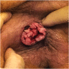
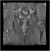
Case presentation: A 45-year-old Caucasian male presented with a perianal mass found to be squamous cell carcinoma. Initial imaging suggested the anal sphincter was spared, however intraoperatively tumor cells were found involving superficial external sphincter fibers and a portion was excised to ensure complete removal.
Clinical discussion: Perianal squamous malignancies are often misdiagnosed as more benign conditions. Treatment aims to preserve sphincter function and depends on tumor stage along with anatomical involvement.
Conclusion: Despite superficial muscle infiltration, the T2N0 perianal lesion was curable with surgical resection alone without recurrence or functional deficits reported one year later. This suggests surgical management may be possible in some cases with sphincter involvement.
Keywords: Anal margin; Case report; General surgery; Oncology; Squamous cell carcinoma.
Copyright © 2021 The Authors. Published by Elsevier Ltd.. All rights reserved.
Figures
References
-
- Gonzalez R.S. Anus & Perianal Area Carcinoma: Squamous Cell Carcinoma. https://www.pathologyoutlines.com/topic/anusscc.html [cited 2021 Feb 2]. Available from:
-
- Agha R.A., Franchi T., Sohrabi C., Mathew G., for the SCARE Group The SCARE 2020 guideline: updating consensus surgical CAse REport (SCARE) guidelines. Int. J. Surg. 2020;84:226–230. - PubMed
Publication types
LinkOut - more resources
Full Text Sources
Other Literature Sources


