. 2021 Jun;112(6):2223-2232.
doi: 10.1111/cas.14894. Epub 2021 Apr 7.

The effect of interleukin-17F on vasculogenic mimicry in oral tongue squamous cell carcinoma

Affiliations

The effect of interleukin-17F on vasculogenic mimicry in oral tongue squamous cell carcinoma

Rabeia Almahmoudi et al. Cancer Sci. 2021 Jun.

Abstract

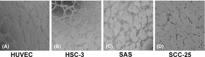
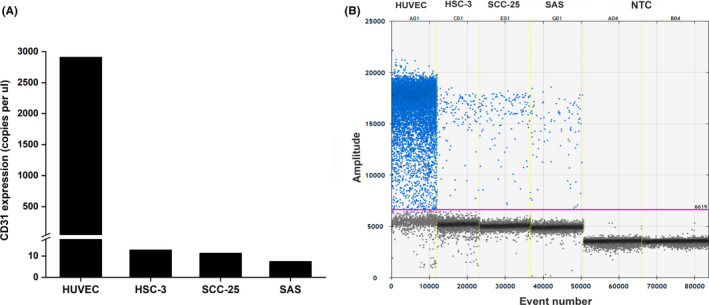
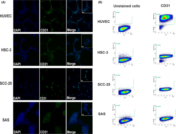
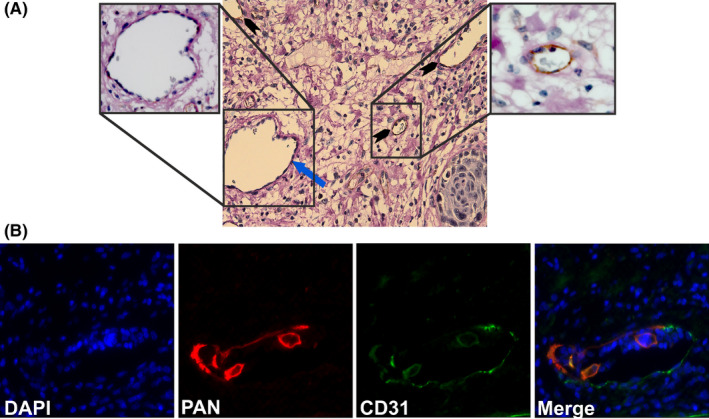
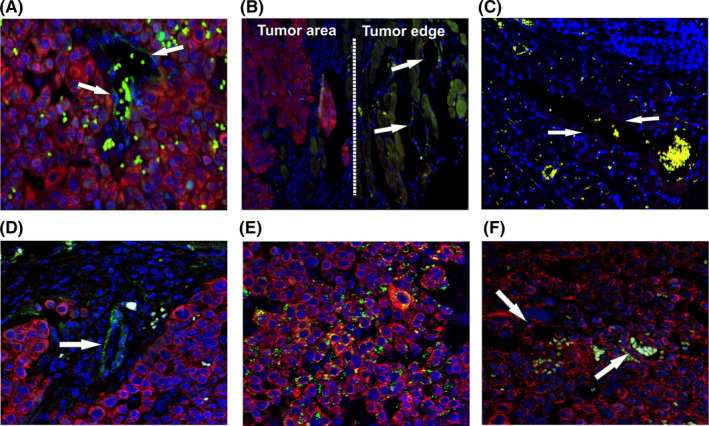
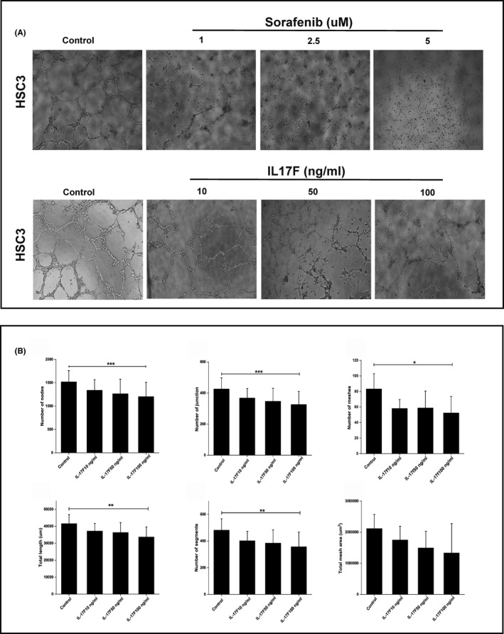
Oral tongue squamous cell carcinoma (OTSCC) is one of the most common cancers worldwide and is characterized by early metastasis and poor prognosis. Recently, we reported that extracellular interleukin-17F (IL-17F) correlates with better disease-specific survival in OTSCC patients and has promising anticancer effects in vitro. Vasculogenic mimicry (VM) is the formation of an alternative vasculogenic system by aggressive tumor cells, which is implicated in treatment failure and poor survival of cancer patients. We sought to confirm the formation of VM in OTSCC and to investigate the effect of IL-17F on VM formation. Here, we showed that highly invasive OTSCC cells (HSC-3 and SAS) form tube-like VM on Matrigel similar to those formed by human umbilical vein endothelial cells. Interestingly, the less invasive cells (SCC-25) did not form any VM structures. Droplet-digital PCR, FACS, and immunofluorescence staining revealed the presence of CD31 mRNA and protein in OTSCC cells. Additionally, in a mouse orthotopic model, HSC-3 cells expressed VE-cadherin (CD144) but lacked Von Willebrand Factor. We identified different patterns of VM structures in patient samples and in an orthotopic OTSCC mouse model. Similar to the effect produced by the antiangiogenic drug sorafenib, IL-17F inhibited the formation of VM structures in vitro by HSC-3 and reduced almost all VM-related parameters. In conclusion, our findings indicate the presence of VM in OTSCC and the antitumorigenic effect of IL-17F through its effect on the VM. Therefore, targeting IL-17F or its regulatory pathways may lead to promising therapeutic strategies in patients with OTSCC.

Keywords: Angiogenesis; CD31; Interleukin-17F; Oral tongue squamous cell carcinoma; Vasculogenic mimicry.


Conflict of interest statement

The authors have no conflict of interest.

Figures

FIGURE 1
FIGURE 1
Cancer cells form tube‐like structures on Matrigel. A‐C, The high‐invasive oral tongue squamous cell carcinoma (OTSCC) cell lines (HSC‐3 and SAS) formed interconnected tube‐like structures that were similar to the structures formed by human umbilical vein endothelial cells (HUVEC) on Matrigel. D, The low‐invasive OTSCC cells (SCC‐25) did not form any consistent tube‐like structures when cultured under the same conditions. Magnification: 4×
FIGURE 2
FIGURE 2
Gene analysis of CD31 in tumor cells. Absolute quantification by droplet‐digital PCR revealed positive expression of CD31 in oral tongue squamous cell carcinoma (OTSCC) cells HSC‐3, SCC‐25, and SAS in vitro (average of approximately 10 copies/µl), which was expectedly less than the control (human umbilical vein endothelial cells; HUVEC)
FIGURE 3
FIGURE 3
Double‐label immunofluorescence and FACS analysis of CD31. A, Cultured oral tongue squamous cell carcinoma (OTSCC) cell lines showed distinct positive staining of CD31. Magnification: 20× and 63×. B, Representative profiles from FACS of OTSCC cells revealed a CD31 expression level of 96% (human umbilical vein endothelial cells; HUVEC), 90% (HSC‐3), 44% (SCC‐25), and 1.8% (SAS)
FIGURE 4
FIGURE 4
Identification of vasculogenic mimicry (VM) structures in oral cancer patients. A, Identification of PAS+/CD31 VM structures. Paraffin‐embedded resection samples from oral tongue squamous cell carcinoma (OTSCC) patients were stained with anti‐human CD31 (brown), periodic acid‐Schiff (PAS: pink), and hematoxylin (purple). PAS+/CD31 vessel‐like structures (blue arrow) and PAS+/CD31+ blood vessels (black arrow heads) were identified. Magnification: 40×. B, Identification of pan‐cytokeratin+/CD31+ mosaic pattern of VM. Pan‐cytokeratin+/CD31+ mosaic structures of VM in OTSCC patients (pan‐cytokeratin: red; CD31: green)
FIGURE 5
FIGURE 5
Identification of VM structures in an orthotopic mouse model of oral cancer. A, Surgical specimens of an orthotopic mouse model of oral tongue squamous cell carcinoma (OTSCC) showed formation of vessels positive for human CD31 (pan‐cytokeratin: red; CD31: green) within and adjacent to the tumor area (arrows). B‐C, Human CD31+ vessels were not found in the adjacent mouse tissue (arrows). D, Confocal microscopy confirmed the presence of CD31+ vessels in vivo. E, Human tumor cells showed abundant staining intensity of CD144 (VE‐cadherin). F, No evident staining pattern of Von Willebrand factor was observed in the orthotopic mouse model of OTSCC (green: red blood cell autofluorescence). Magnification: 40×
FIGURE 6
FIGURE 6
A, Interleukin‐17F inhibits formation of vasculogenic mimicry (VM) in vitro. The effect of IL‐17F on VM formation on Matrigel was compared with the angiogenesis inhibitor sorafenib. When treated with sorafenib for 24 h, HSC‐3 cells showed reduction in tube formation at concentrations of 1‐5 µM. Similarly, IL‐17F inhibited the formation of the tube‐like structures in HSC‐3 cells compared with control, with the greatest effect observed at 100 ng/ml. Magnification: 4×. B, Interleukin‐17F reduces the parameters of VM in vitro. A tube formation analysis tool was used to further analyze the IL‐17F effect on VM parameters. IL‐17F at 100 ng/ml reduced almost all VM‐related parameters in HSC‐3, including number of nodes (P =.001), number of junctions (P =.001), number of meshes (P =.02), total length (P =.004), and number of segments (P =.009). Total mesh areas were also reduced by IL‐17F, but the difference was not significant compared with the controls. Experiments were performed independently at least three times. Values are expressed as means ± SD. Mann‐Whitney U test and Friedman test were performed on independent samples

Similar articles

Cited by

References

    1. Miranda‐Filho A, Bray F. Global patterns and trends in cancers of the lip, tongue and mouth. Oral Oncol. 2020;24(102):104551. 10.1016/j.oraloncology.2019.104551 - DOI - PubMed
    1. Kim YJ, Kim JH. Increasing incidence and improving survival of oral tongue squamous cell carcinoma. Sci Rep. 2020;10(1):7877. 10.1038/s41598-020-64748-0 - DOI - PMC - PubMed
    1. Ng JH, Iyer NG, Tan MH, Edgren G. Changing epidemiology of oral squamous cell carcinoma of the tongue: a global study. Head Neck. 2016;39:297‐304. - PubMed
    1. Van Dijk BA, Brands MT, Geurts SM, Merkx MA, Roodenburg JL. Trends in oral cavity cancer incidence, mortality, survival and treatment in the Netherlands. Int J Cancer. 2016;139:574‐583. - PubMed
    1. da Silva SD, Hier M, Mlynarek A, Kowalski LP, Alaoui‐Jamali MA. Recurrent oral cancer: current and emerging therapeutic approaches. Front Pharmacol. 2012;30:149. - PMC - PubMed

MeSH terms