Case Reports
. 2021 Mar 20;21(1):55.
doi: 10.1186/s12880-021-00585-5.

A 78-year-old female with severe tongue pain: benefit of modern ultrasound

Affiliations
Case Reports

A 78-year-old female with severe tongue pain: benefit of modern ultrasound

Lara Clarissa Burg et al. BMC Med Imaging. .

Abstract

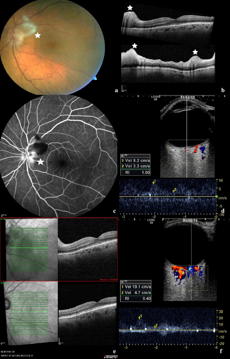
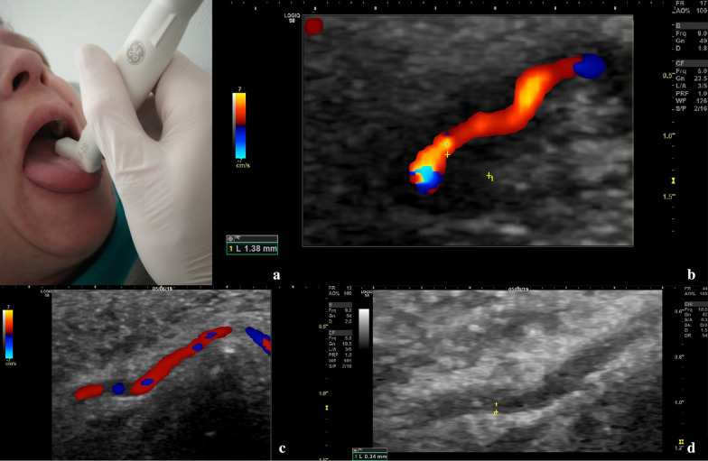
Background: Giant cell arteritis (GCA) is the most common form of systemic vasculitis in persons aged 50 years and older. Medium and large vessels, like the temporal and axillary arteries, are commonly affected. Typical symptoms are headache, scalp tenderness, jaw claudication and ophthalmological symptoms as loss of visual field, diplopia or amaurosis due to optic nerve ischemia. Tongue pain due to vasculitic affection of the deep lingual artery can occur and has so far not been visualized and followed up by modern ultrasound.

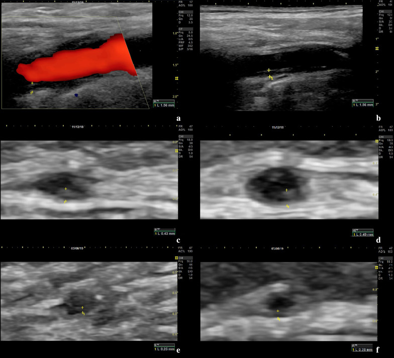
Case presentation: We report the case of a 78-year-old woman with typical symptoms of GCA, such as scalp tenderness, jaw claudication and loss of visual field, as well as severe tongue pain. Broad vasculitic affection of the extracranial arteries, vasculitis of the central retinal artery and the deep lingual artery could be visualized by ultrasound. Further did we observe a relevant decrease of intima-media thickness (IMT) values of all arteries assessed by ultrasound during follow-up. Especially the left common superficial temporal artery showed a relevant decrease of IMT from 0.49 mm at time of diagnosis to 0.23 mm on 6-months follow-up. This is the first GCA case described in literature, in which vasculitis of the central retinal artery and the lingual artery could be visualized at diagnosis and during follow-up using high-resolution ultrasound.

Conclusion: High-resolution ultrasound can be a useful diagnostic imaging modality in diagnosis and follow-up of GCA, even in small arteries like the lingual artery or central retinal artery. Ultrasound of the central retinal artery could be an important imaging tool in identifying suspected vasculitic affection of the central retinal artery.

Keywords: Central retinal artery; Follow-up; Giant cell arteritis; Intima-media thickness; Lingual artery; Ultrasound; Vasculitis.


Conflict of interest statement

The authors declare that they have no competing interests.

Figures

Fig. 1
Fig. 1
Ophthalmologic imaging and transocular ultrasound of the left eye. a Colour fundus retinal photography of the left eye displaying an optic disc edema with adjacent cotton wool spot (*) along the upper temporal vascular arch. b Macula-optical coherence tomography of the left eye with a normal macula and segmental disc edema. Retinal nerve fibre layer thickness was increased in the superior, temporal and inferior segments (*). c Fluorescein angiography of the left eye with distinct hyperfluorescence of the papilla, i.e. a “hot disc”. d Transocular ultrasound including measurement of flow velocity of the central retinal artery of the left eye on admission. e Macula-optical coherence tomography on admission (top) compared to 6-month follow-up (bottom). f Transocular ultrasound including measurement of flow velocity of the central retinal artery of the left eye in 6-month follow-up
Fig. 2
Fig. 2
Ultrasound images displaying vasculitic changes of the intima-media complex of the respective arteries. a, b Left vertebral artery in Doppler and B-mode ultrasound with pathological vessel wall and increased intima-media thickness (IMT) due to inflammation. c Right common superficial temporal artery with pathological IMT of 0.43 mm. d Left common superficial temporal artery with pathological IMT of 0.49 mm. e Right common superficial temporal artery in 6-month follow-up with IMT of 0.25 mm. f Left common superficial temporal artery in 6-month follow-up with IMT of 0.23 mm
Fig. 3
Fig. 3
Ultrasound examination of the lingual artery with an 18 MHz hockey stick probe. a Position of the ultrasound probe on the patient´s tongue. b Doppler ultrasound and intima-media thickness (IMT) values of the affected deep lingual artery. c Doppler ultrasound of the deep lingual artery on 6-month follow-up, reduced field of view. d IMT values of the deep lingual artery on 6-month follow-up in B-Mode, reduced field of view

References

    1. Warrington KJ, Matteson EL. Management guidelines and outcome measures in giant cell arteritis (GCA) Clin Exp Rheumatol. 2007;25:137–141. - PubMed
    1. Truffaut L, Lefebvre P. Tongue necrosis in giant-cell arteritis. N Engl J Med. 2018;378:2517. doi: 10.1056/NEJMicm1709412. - DOI - PubMed
    1. Grant SWJ, Underhill HC, Atkin P. Giant cell arteritis affecting the tongue: a case report and review of the literature. Dent Update. 2013;40:669–670. doi: 10.12968/denu.2013.40.8.669. - DOI - PubMed
    1. Schäfer VS, Chrysidis S, Dejaco C, Duftner C, Iagnocco A, Bruyn GA, et al. Assessing vasculitis in giant cell arteritis by ultrasound: results of OMERACT patient-based reliability exercises. J Rheumatol. 2018;45:1289–1295. doi: 10.3899/jrheum.171428. - DOI - PubMed
    1. Schmidt W, Kraft H, Völker L, Vorpahl K, Gromnica-Ihle E. Colour doppler sonography to diagnose temporal arteritis. Lancet. 1995;345:866. doi: 10.1016/S0140-6736(95)93005-1. - DOI - PubMed

Publication types

LinkOut - more resources