. 2021 Mar 20;19(1):81.
doi: 10.1186/s12951-021-00827-2.

Biomimetic oxygen delivery nanoparticles for enhancing photodynamic therapy in triple-negative breast cancer

Affiliations

Biomimetic oxygen delivery nanoparticles for enhancing photodynamic therapy in triple-negative breast cancer

Hanyi Fang et al. J Nanobiotechnology. .

Abstract

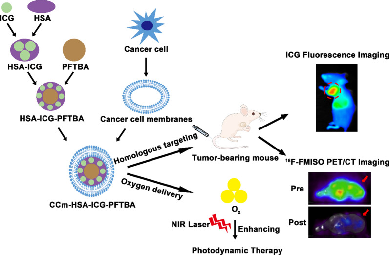
Background: Triple-negative breast cancer (TNBC) is a kind of aggressive breast cancer with a high rate of metastasis, poor overall survival time, and a low response to targeted therapies. To improve the therapeutic efficacy and overcome the drug resistance of TNBC treatments, here we developed the cancer cell membrane-coated oxygen delivery nanoprobe, CCm-HSA-ICG-PFTBA, which can improve the hypoxia at tumor sites and enhance the therapeutic efficacy of the photodynamic therapy (PDT), resulting in relieving the tumor growth in TNBC xenografts.

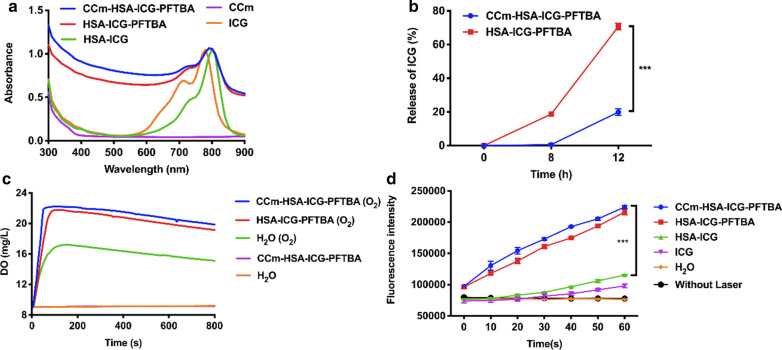
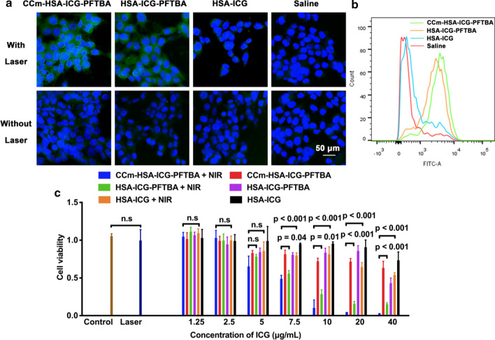
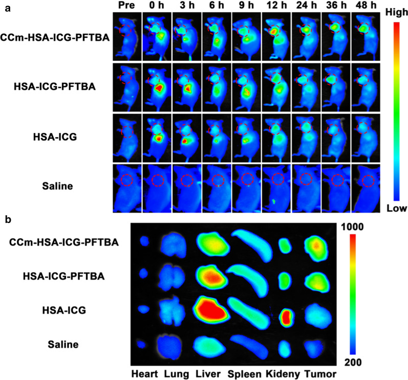
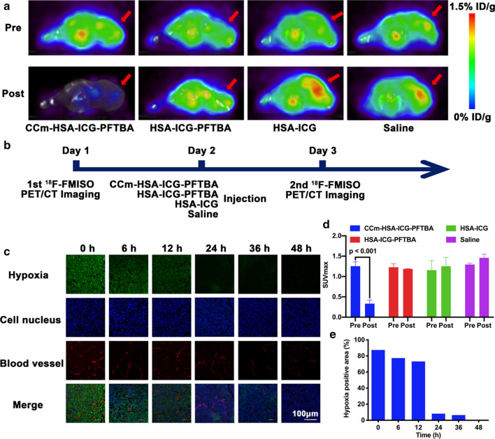
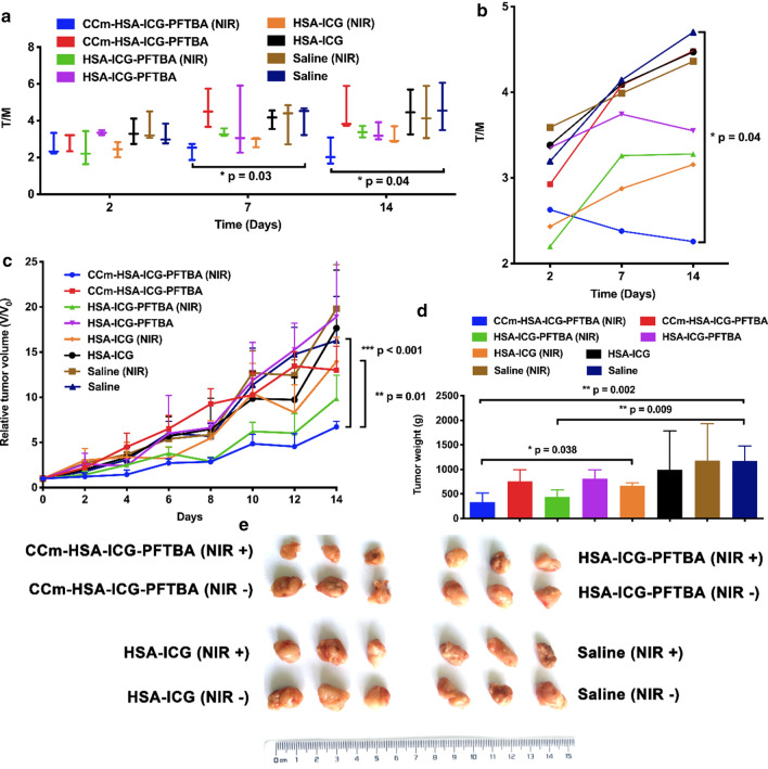
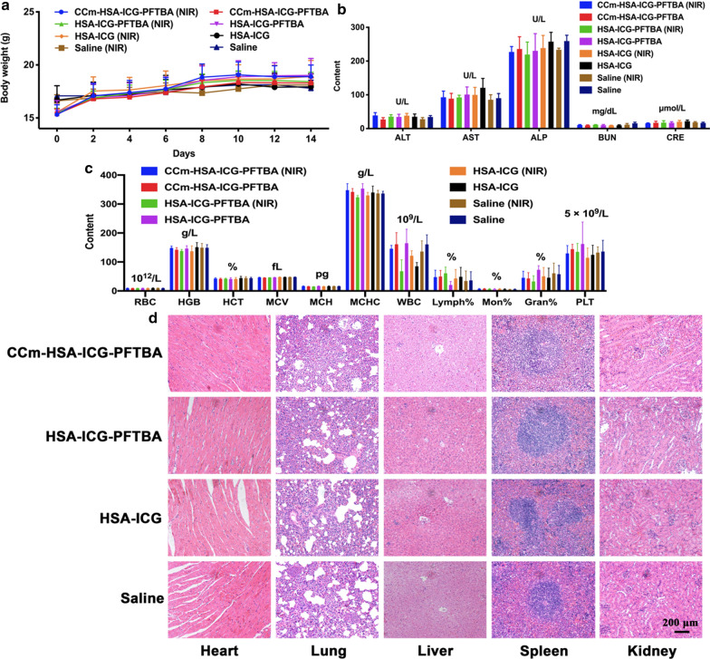
Results: The size of the CCm-HSA-ICG-PFTBA was 131.3 ± 1.08 nm. The in vitro 1O2 and ROS concentrations of the CCm-HSA-ICG-PFTBA group were both significantly higher than those of the other groups (P < 0.001). In vivo fluorescence imaging revealed that the best time window was at 24 h post-injection of the CCm-HSA-ICG-PFTBA. Both in vivo 18F-FMISO PET imaging and ex vivo immunofluorescence staining results exhibited that the tumor hypoxia was significantly improved at 24 h post-injection of the CCm-HSA-ICG-PFTBA. For in vivo PDT treatment, the tumor volume and weight of the CCm-HSA-ICG-PFTBA with NIR group were both the smallest among all the groups and significantly decreased compared to the untreated group (P < 0.01). No obvious biotoxicity was observed by the injection of CCm-HSA-ICG-PFTBA till 14 days.

Conclusions: By using the high oxygen solubility of perfluorocarbon (PFC) and the homologous targeting ability of cancer cell membranes, CCm-HSA-ICG-PFTBA can target tumor tissues, mitigate the hypoxia of the tumor microenvironment, and enhance the PDT efficacy in TNBC xenografts. Furthermore, the HSA, ICG, and PFC are all FDA-approved materials, which render the nanoparticles highly biocompatible and enhance the potential for clinical translation in the treatment of TNBC patients.

Keywords: Cancer cell membranes; Hypoxia; Nanoprobes; Oxygen delivery; Photodynamic therapy; Triple-negative breast cancer.


Conflict of interest statement

The authors declare no competing interests.

Figures

Scheme 1.
Scheme 1.
Illustration of the biomimetic oxygen-delivery nanoprobe. It was cancer cell membrane-coated indocyanine green-doped perfluorocarbon (CCm–HSA–ICG–PFTBA) for homologous targeting and improving oxygen concentration at tumor sites. 18F-FMISO PET/CT imaging was performed to measure the hypoxia in vivo. CCm–HSA–ICG–PFTBA was injected into 4T1 xenografts and then photodynamic therapy was performed. Tumor volume was measured to evaluate the therapeutic efficacy enhancement
Fig. 1
Fig. 1
Characterization of CCm–HSA–ICG–PFTBA. a Size intensity curves, b hydrodynamic size, c Zeta potential of CCm–HSA–ICG–PFTBA, HSA–ICG–PFTBA, and cancer cell membrane (CCm). d Stability of CCm–HSA–ICG–PFTBA and HSA–ICG–PFTBA. ei TEM images of eh CCm–HSA–ICG–PFTBA, fi HSA–ICG–PFTBA, and g CCm. Scale bars = 30 nm and 50 nm. Data are represented as mean ± SD (n = 3)
Fig. 2
Fig. 2
Properties of CCm–HSA–ICG–PFTBA. a UV–vis absorbance spectra of CCm–HSA–ICG–PFTBA, HSA–ICG–PFTBA, HSA–ICG, CCm, and ICG. b The release of CCm–HSA–ICG–PFTBA and HSA–ICG–PFTBA in serum under 37 ℃. c Oxygen concentrations in water after adding CCm–HSA–ICG–PFTBA and HSA–ICG–PFTBA with or without pre-oxygenation. d Enhanced 1O2 generation of CCm–HSA–ICG–PFTBA, HSA–ICG–PFTBA, HSA–ICG, and ICG. Data are represented as mean ± SD (n = 3). *** indicates P < 0.001
Fig. 3
Fig. 3
In vitro experiments of CCm–HSA–ICG–PFTBA. a The confocal laser scanning microscope images of ROS generation in cells treated with the CCm–HSA–ICG–PFTBA, HSA–ICG–PFTBA, HSA–ICG, and saline with or without NIR laser irradiation. Green fluorescence represented DCFH-DA, showing ROS concentrations, and blue fluorescence represented DAPI, showing cell nucleus. The scale bar = 50 μm. b Flow cytometry analysis of ROS generation in cells treated with the CCm–HSA–ICG–PFTBA, HSA–ICG–PFTBA, HSA–ICG, and saline with NIR laser irradiation. c Cell viability after treated with the CCm–HSA–ICG–PFTBA, HSA–ICG–PFTBA, and HSA–ICG with or without NIR laser irradiation. Data are represented as mean ± SD (n = 5)
Fig. 4
Fig. 4
Fluorescence imaging. a In vivo fluorescence images of TNBC xenografts after the injection of the CCm–HSA–ICG–PFTBA, HSA–ICG–PFTBA, HSA–ICG, and saline at different time points. Red circles indicated the tumor site. b Ex vivo fluorescence images of major organs and tumors at 48 h post-injection
Fig. 5
Fig. 5
Hypoxia improvement at tumor sites. a In vivo transverse 18F-FMISO PET/CT images of TNBC xenografts before and after 24 h injection of the CCm–HSA–ICG–PFTBA, HSA–ICG–PFTBA, HSA–ICG, and saline. Red arrows indicated tumor sites. b Scheme of the PET/CT imaging. c Immunofluorescence images of tumor slices stained by the hypoxyprobe. The blood vessels and hypoxia areas were stained with anti-CD31 antibody (red) and antipimonidazole antibody (green), respectively. Scale bar = 150 μm. d The quantitative analysis of SUVmax at tumor sites of CCm–HSA–ICG–PFTBA, HSA–ICG–PFTBA, HSA–ICG, and saline groups in the pre and post 18F-FMISO PET/CT imaging. e Quantification of tumor hypoxia densities for different time points
Fig. 6
Fig. 6
In vivo PDT of tumor-bearing mice. a, b The tumor to muscle (T/M) ratios of mice after treated with CCm–HSA–ICG–PFTBA, HSA–ICG–PFTBA, HSA–ICG, and saline and with or without NIR laser irradiation. T/M was calculated by drawn ROI in the images of 18F-FDG PET images. Values are the means ± SD (n = 3). c Relative tumor volume, d the tumor weight obtained after treatment for 14 days, and e representative photographs of tumor tissues of the mice in different treatment groups. Data are represented as mean ± SD (n = 6). *, **, and *** indicate P < 0.05, P < 0.01 and P < 0.001, respectively
Fig. 7
Fig. 7
In vivo toxicity evaluation. a Mice body-weight-change curves over 14 days after injection with CCm–HSA–ICG–PFTBA, HSA–ICG–PFTBA, HSA–ICG, and saline, with or without NIR irradiation. b Blood parameters data. Red blood cells (RBC), hemoglobin (HGB), hematocrit (HCT), mean corpuscular volume (MCV), mean corpuscular hemoglobin (MCH), and mean corpuscular hemoglobin concentration (MCHC). c Blood biochemistry data. Alanine transaminase (ALT), aspartate aminotransferase (AST), alkaline phosphatase (ALP), blood urea nitrogen (BUN), and creatinine (CRE). d White blood cells (WBC), lymphocytes percentage (Lymph%), monocyte percentage (Mon%), and neutrophil percentage (Neu%). e Platelets (PLT). f H&E-stained slice images of major organs. Scale bars = 200 μm. Data are represented as mean ± SD (n = 6)

References

    1. Siegel RL, Miller KD, Jemal A. Cancer statistics, 2020. CA Cancer J Clin. 2020;70:7–30. doi: 10.3322/caac.21590. - DOI - PubMed
    1. Waks AG, Winer EP. Breast cancer treatment: a review. JAMA. 2019;321:288–300. doi: 10.1001/jama.2018.19323. - DOI - PubMed
    1. Bianchini G, Balko JM, Mayer IA, Sanders ME, Gianni L. Triple-negative breast cancer: challenges and opportunities of a heterogeneous disease. Nat Rev Clin Oncol. 2016;13:674–690. doi: 10.1038/nrclinonc.2016.66. - DOI - PMC - PubMed
    1. Medina MA, Oza G, Sharma A, Arriaga LG, Hernandez Hernandez JM, Rotello VM, Ramirez JT. Triple-negative breast cancer: a review of conventional and advanced therapeutic strategies. Int J Environ Res Public Health. 2020;17:2078. doi: 10.3390/ijerph17062078. - DOI - PMC - PubMed
    1. Agostinis P, Berg K, Cengel KA, Foster TH, Girotti AW, Gollnick SO, Hahn SM, Hamblin MR, Juzeniene A, Kessel D, et al. Photodynamic therapy of cancer: an update. CA Cancer J Clin. 2011;61:250–281. doi: 10.3322/caac.20114. - DOI - PMC - PubMed

MeSH terms

LinkOut - more resources