. 2021 Mar 20;12(1):199.
doi: 10.1186/s13287-021-02275-z.

Human placental mesenchymal stem cells ameliorate chemotherapy-induced damage in the testis by reducing apoptosis/oxidative stress and promoting autophagy

Affiliations

Human placental mesenchymal stem cells ameliorate chemotherapy-induced damage in the testis by reducing apoptosis/oxidative stress and promoting autophagy

Jiafeng Lu et al. Stem Cell Res Ther. .

Abstract

Background: The side effects of busulfan on male reproduction are serious, so fertility preservation in children undergoing busulfan treatment is a major worldwide concern. Human placental mesenchymal stem cells (hPMSCs) have advantages such as stable proliferation and lower immunogenicity that make them an ideal material for stimulating tissue repair, especially restoring spermatogenesis. The protective effects of hPMSCs in busulfan-induced Sertoli cells and in busulfan-treated mouse testes have not been determined. Our study aimed to elaborate the protective effect and potential mechanisms of hPMSCs in busulfan-treated testes and Sertoli cells.

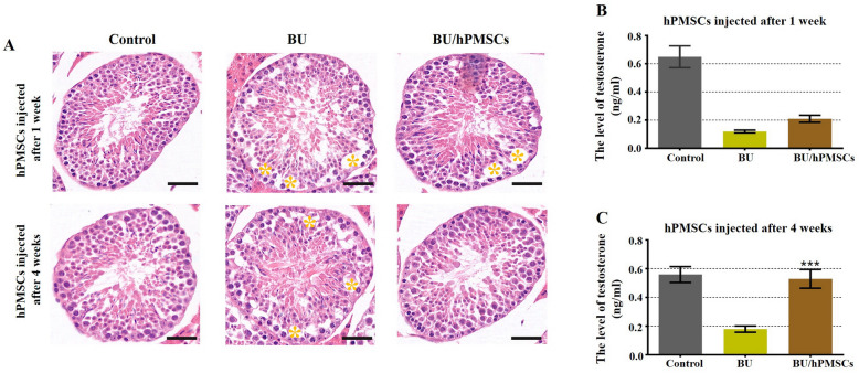
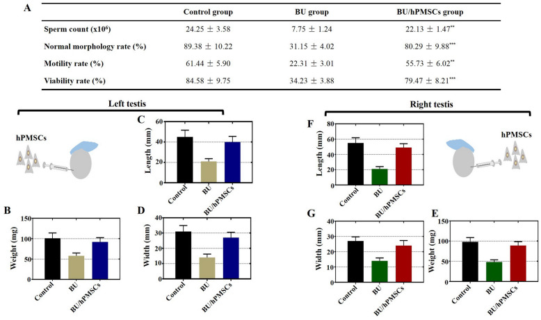
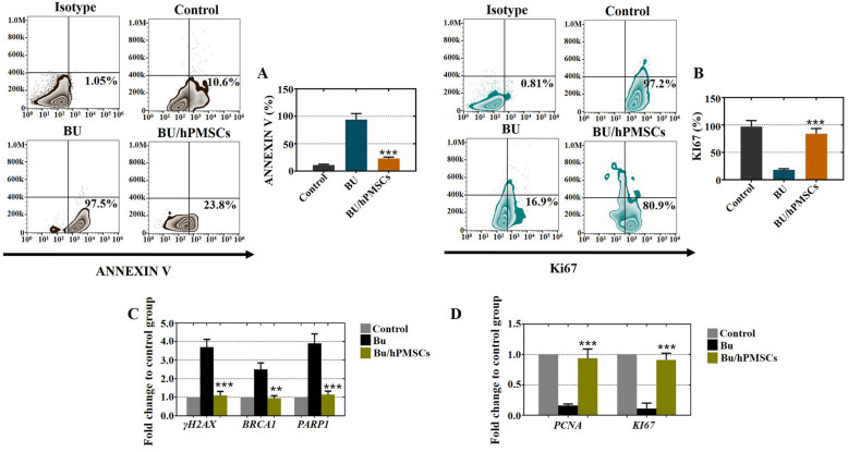
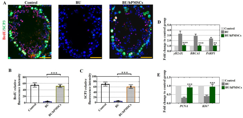
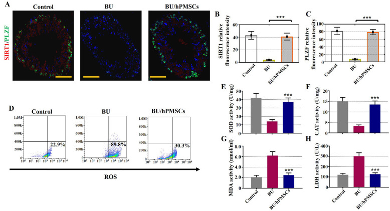
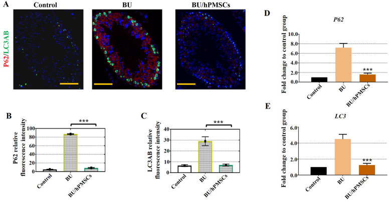
Methods: First, we developed a mouse model of busulfan-induced testicular toxicity in vivo and a mouse Sertoli cell line treated with busulfan in vitro to assess the protective effect and mechanisms of hPMSC treatment on spermatogenesis. Then, the length, width, and weight of the testes were monitored using Vernier calipers. Furthermore, at 1 week and 4 weeks after the transplantation of hPMSCs, histological sections of testes were stained with hematoxylin-eosin, and the seminiferous tubules with fluid-filled cavities were counted. Through ELISA analysis, testosterone levels and MDA, SOD, LDH, and CAT activities, which are associated with ROS, were detected. Markers of ROS, proliferation (Ki67), and apoptosis (Annexin V) were evaluated by FACS. Next, the fluorescence intensity of proliferation markers (BrdU and SCP3), an antioxidant marker (SIRT1), a spermatogenesis marker (PLZF), and autophagy-related genes (P62 and LC3AB) were detected by fluorescence microscopy. The mRNA expression of γ-H2AX, BRCA1, PARP1, PCNA, Ki67, P62, and LC3 was determined by qRT-PCR.

Results: hPMSCs restored disrupted spermatogenesis, promoted improved semen parameters, and increased testosterone levels, testis size, and autophagy in the testis toxicity mouse model induced by busulfan. hPMSCs suppressed the apoptosis of Sertoli cells and enhanced their rate of proliferation in vitro. Additionally, hPMSCs protected against oxidative stress and decreased oxidative damage in the testis toxicity mouse model induced by busulfan. Furthermore, hPMSCs increased the expression of proliferation genes (PCNA and KI67) and decreased the mRNA levels of apoptotic genes such as γ-H2AX, BRCA1, and PARP1.

Conclusions: This research showed that hPMSC injection ameliorated busulfan-induced damage in the testis by reducing apoptosis/oxidative stress and promoting autophagy. The present study offers an idea for a new method for clinical treatment of chemotherapy-induced spermatogenesis.

Keywords: Apoptosis; Autophagy; Busulfan; Human placental mesenchymal stem cells; Reactive oxygen species; Spermatogenesis.


Conflict of interest statement

The authors declare no conflicts of interest.

Figures

Fig. 1
Fig. 1
Human placental mesenchymal stem cells (hPMSCs) rescued spermatogenesis and promoted testosterone levels in a busulfan-induced testis toxicity mouse model. a Images of HE-stained mouse testis sections in the three groups (the control group and the 1- and 4-week hPMSC treatment groups). Scale bar = 20 μm. n = 3 for each group. b Testosterone levels were examined 1 week after hPMSC injection in the three groups. c Testosterone levels were detected 4 weeks after hPMSC treatment in the three groups. The error bars indicate the SD. ***p < 0.001
Fig. 2
Fig. 2
hPMSCs promoted increased testicular weight and size and improved semen parameters in a busulfan-induced testis toxicity mouse model. a The sperm count, normal morphology, motility, and proportion of viable cells were determined in the three groups after hPMSC therapy. bd Variation in the weight, length, and width of the mouse left testis in the three groups after hPMSC treatment. eg Variation in the weight, length, and width of the mouse right testis in the three groups after hPMSC treatment. The error bars indicate the SD. **p < 0.01,***p < 0.001
Fig. 3
Fig. 3
In cultured Sertoli cells treated with busulfan, hPMSCs inhibited cell apoptosis and enhanced cell proliferation. a The FACS results indicated that hPMSC treatment inhibited the rate of apoptosis (Annexin V) in Sertoli cells. b The FACS results showed that hPMSC treatment improved the proliferation rate (Ki67) of Sertoli cells. c, d The qRT-PCR results showed that hPMSC treatment suppressed the expression of apoptotic genes (γ-H2AX, BRCA1, and PARP1) and increased the expression of proliferative genes (PCNA and Ki67). All experiments were carried out three times, and the error bars indicate the SD. **p < 0.01, ***p < 0.001
Fig. 4
Fig. 4
hPMSCs improved cell proliferation and alleviated apoptosis of testes in the busulfan-induced testis toxicity mouse model. ac The fluorescence intensities of BrdU (red) and SCP3 (green) in the three groups were detected by fluorescence microscopy. d, e The mRNA expression of γ-H2AX, BRCA1, PARP1, PCNA, and Ki67 was determined by qRT-PCR. The error bars indicate the SD. **p < 0.01, ***p < 0.001
Fig. 5
Fig. 5
hPMSCs alleviated oxidative damage in a busulfan-induced testis toxicity mouse model. ac Fluorescence microscopy was used to detect the relative fluorescence intensity of SIRT1 (red) and PLZF (green) in the three groups. d FACS analysis was applied to measure the percentage of ROS+ Sertoli cells in the three groups. eh ELISA analysis was employed to examine the expression of SOD, CAT, MDA, and LDH. The error bars indicate the SD. ***p < 0.001
Fig. 6
Fig. 6
hPMSCs promoted autophagy in a busulfan-induced testis toxicity mouse model. ac Fluorescence microscopy was employed to monitor the relative fluorescence intensities of p62 (red) and LC3AB (green) in the three groups. de qRT-PCR assays revealed the mRNA levels of the autophagy-related genes P62 and LC3. The error bars indicate the SD. ***p < 0.001

References

    1. Nishimura H, L’Hernault SW. Spermatogenesis. Curr Biol. 2017;27(18):R988–R994. doi: 10.1016/j.cub.2017.07.067. - DOI - PubMed
    1. Hai Y, Hou J, Liu Y, Liu Y, Yang H, Li Z, He Z. The roles and regulation of Sertoli cells in fate determinations of spermatogonial stem cells and spermatogenesis. Semin Cell Dev Biol. 2014;29:66–75. doi: 10.1016/j.semcdb.2014.04.007. - DOI - PubMed
    1. Matzuk MM, Lamb DJ. The biology of infertility: research advances and clinical challenges. Nat Med. 2008;14(11):1197–1213. doi: 10.1038/nm.f.1895. - DOI - PMC - PubMed
    1. Zhou Q, Wang M, Yuan Y, Wang X, Fu R, Wan H, Xie M, Liu M, Guo X, Zheng Y, Feng G, Shi Q, Zhao XY, Sha J, Zhou Q. Complete meiosis from embryonic stem cell-derived germ cells in vitro. Cell Stem Cell. 2016;18(3):330–340. doi: 10.1016/j.stem.2016.01.017. - DOI - PubMed
    1. Levine H, Jørgensen N, Martino-Andrade A, Mendiola J, Weksler-Derri D, Mindlis I, Pinotti R, Swan SH. Temporal trends in sperm count: a systematic review and meta-regression analysis. Hum Reprod Update. 2017;23(6):646–659. doi: 10.1093/humupd/dmx022. - DOI - PMC - PubMed

Publication types

Substances