Chest computed tomography findings in different phases of SARS-CoV-2 infection
- PMID: 33743999
- PMCID: PMC8017560
- DOI: 10.1016/j.rx.2021.02.004
Chest computed tomography findings in different phases of SARS-CoV-2 infection
Abstract
Objective: To compare the findings on chest computed tomography (CT) in patients with COVID-19 during different phases of the disease and to evaluate the reproducibility of a visual radiologic score for estimating the extent of lung involvement.
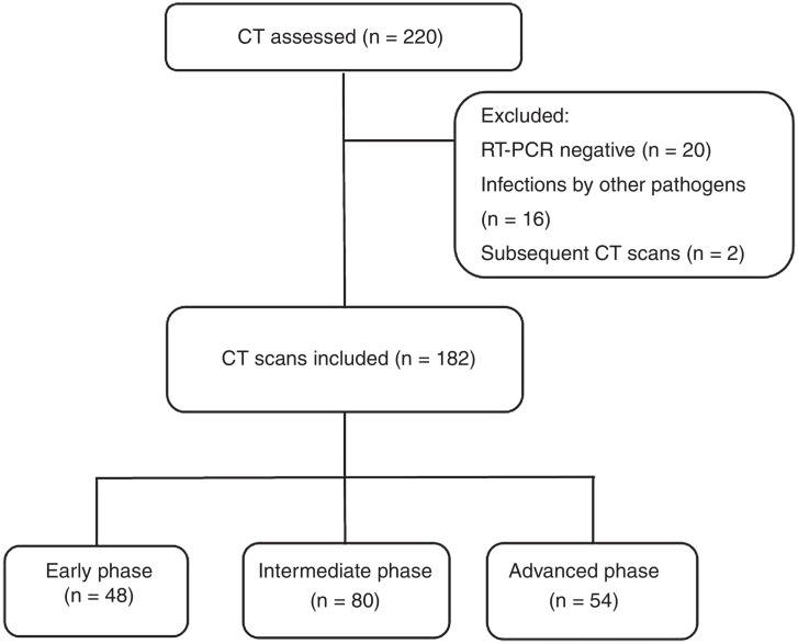
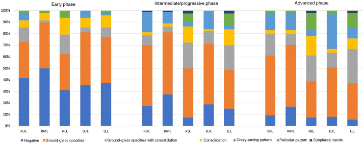
Methods: We retrospectively reviewed chest CT studies from 182 patients with RT-PCR findings positive for SARS-CoV-2. Patients were classified according to the time elapsed from the onset of symptoms, as follows: early (0-4 days), intermediate/progressive (5-9 days), or advanced (≥10 days). We analyzed the frequency of each radiologic finding, as well as the pattern, appearance, and predominant distribution of lung involvement. A visual tomographic score (range, 0-25) was used to estimate the extent of involvement in each lobe and in the total lung volume.
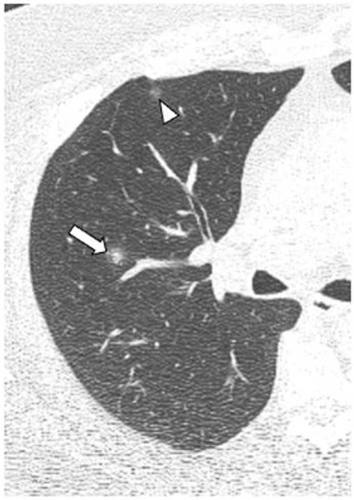
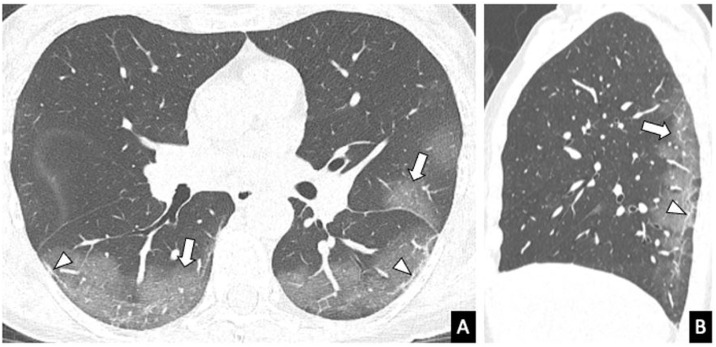
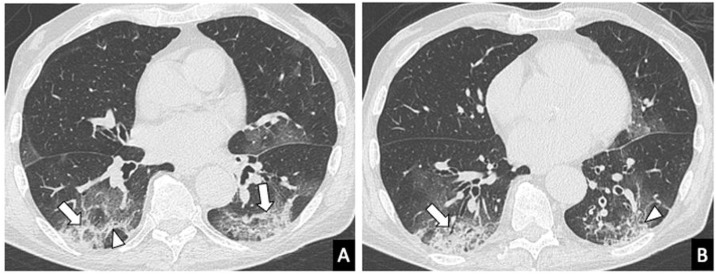
Results: The predominant CT finding was the ground-glass pattern (n=110; 60.4%), the most common distribution was peripheral (n = 116; 66.7%), and the most prevalent appearance was typical (n=112; 61.5%). The halo sign was seen most frequently in the early phase (25%), whereas ground-glass opacities were more common in the intermediate/progressive and advanced phases. The median severity score was 10 (IQR: 5-13), and the scores increased as the disease progressed. The interobserver agreement (kappa) was 0.92 for the appearance, 0.84 for the distribution, 0.70 for the predominant pattern, and 0.89 for the visual score.
Conclusion: The CT findings in patients with COVID-19 vary with the course of the infection. The proposed visual radiologic score is a simple, reproducible, and reliable tool for assessing lung involvement in COVID-19 pneumonia.
Objetivo: Comparar los hallazgos radiológicos mediante tomografía computarizada (TC) torácica en pacientes con COVID-19 en diferentes fases de la enfermedad y evaluar la reproducibilidad de unscore radiológico visual para estimar la extensión de la afectación pulmonar.
Métodos: Se evaluaron retrospectivamente las tomografías computarizadas de tórax de 182 pacientes con RT-PCR positiva para SARS-CoV-2. En función del tiempo de evolución de la infección, los pacientes fueron clasificados en tres grupos/estadios: fase precoz (0–4 días), intermedia/progresiva (5–9 días) y avanzada (≥10 días). Se analizó la frecuencia de cada hallazgo radiológico, así como el patrón, la apariencia y la distribución predominantes de la afectación pulmonar. La extensión de la afectación pulmonar se estimó para cada lóbulo pulmonar y para el volumen pulmonar total mediante un score tomográfico visual (rango 0−25).
Resultados: El hallazgo tomográfico predominante fue el patrón en vidrio deslustrado (n = 110, 60,4%), la distribución más frecuente, la periférica (n = 116, 66,7%) y la apariencia más prevalente, la típica (n = 112, 61,5%). El “signo del halo” se encontró más frecuentemente en la fase precoz (25%), mientras que las opacidades en vidrio deslustrado, el patrón en empedrado y las líneas subpleurales fueron más frecuentes en las fases intermedia/progresiva y avanzada. La mediana delscore de gravedad fue de 10 (RIQ: 5–13), aumentando los valores con la progresión de la enfermedad. El acuerdo interobservador (kappa, k) para la apariencia, la distribución y el patrón predominante, así como para el score visual fueron de 0,92; 0,84; 0,70, y 0,89; respectivamente.
Conclusión: Los hallazgos tomográficos en la COVID-19 varían con el curso de la infección. Elscore radiológico sugerido es una herramienta sencilla, reproducible y fiable para evaluar la afectación pulmonar en la neumonía COVID-19.
Keywords: COVID-19; COVID-19.; Computed tomography; Neumonía; Tomografía computarizada; pneumonia.
Copyright © 2021 SERAM. Publicado por Elsevier España, S.L.U. All rights reserved.
Figures
Similar articles
-
[Chest computed tomography findings in different phases of SARS-CoV-2 infection].Radiologia. 2021 May-Jun;63(3):218-227. doi: 10.1016/j.rx.2021.02.004. Epub 2021 Feb 27. Radiologia. 2021. PMID: 35370313 Free PMC article. Spanish.
-
CT-scan findings of COVID-19 pneumonia based on the time elapsed from the beginning of symptoms to the CT imaging evaluation: a descriptive study in Iran.Rom J Intern Med. 2020 Dec 17;58(4):242-250. doi: 10.2478/rjim-2020-0019. Print 2020 Dec 1. Rom J Intern Med. 2020. PMID: 32726296
-
Frequency and Distribution of Chest Radiographic Findings in Patients Positive for COVID-19.Radiology. 2020 Aug;296(2):E72-E78. doi: 10.1148/radiol.2020201160. Epub 2020 Mar 27. Radiology. 2020. PMID: 32216717 Free PMC article.
-
Predictors of the chest CT score in COVID-19 patients: a cross-sectional study.Virol J. 2021 Nov 18;18(1):225. doi: 10.1186/s12985-021-01699-6. Virol J. 2021. PMID: 34794467 Free PMC article. Review.
-
Comparison of the computed tomography findings in COVID-19 and other viral pneumonia in immunocompetent adults: a systematic review and meta-analysis.Eur Radiol. 2020 Dec;30(12):6485-6496. doi: 10.1007/s00330-020-07018-x. Epub 2020 Jun 27. Eur Radiol. 2020. PMID: 32594211 Free PMC article.
References
-
- Coronavirus disease 2019. World Health Organization. https://covid19.who.int/ (Accessed: 19 January 2021).
-
- Kivu N. 2020. WHO Director-General’s opening remarks at the media briefing on; pp. 1–5.
Publication types
MeSH terms
LinkOut - more resources
Full Text Sources
Other Literature Sources
Medical
Miscellaneous