. 2021 Jan-Jun:296:100551.
doi: 10.1016/j.jbc.2021.100551. Epub 2021 Mar 17.

Glucocorticoid receptor Thr524 phosphorylation by MINK1 induces interactions with 14-3-3 protein regulators

Affiliations

Glucocorticoid receptor Thr524 phosphorylation by MINK1 induces interactions with 14-3-3 protein regulators

Claire C Munier et al. J Biol Chem. 2021 Jan-Jun.

Abstract

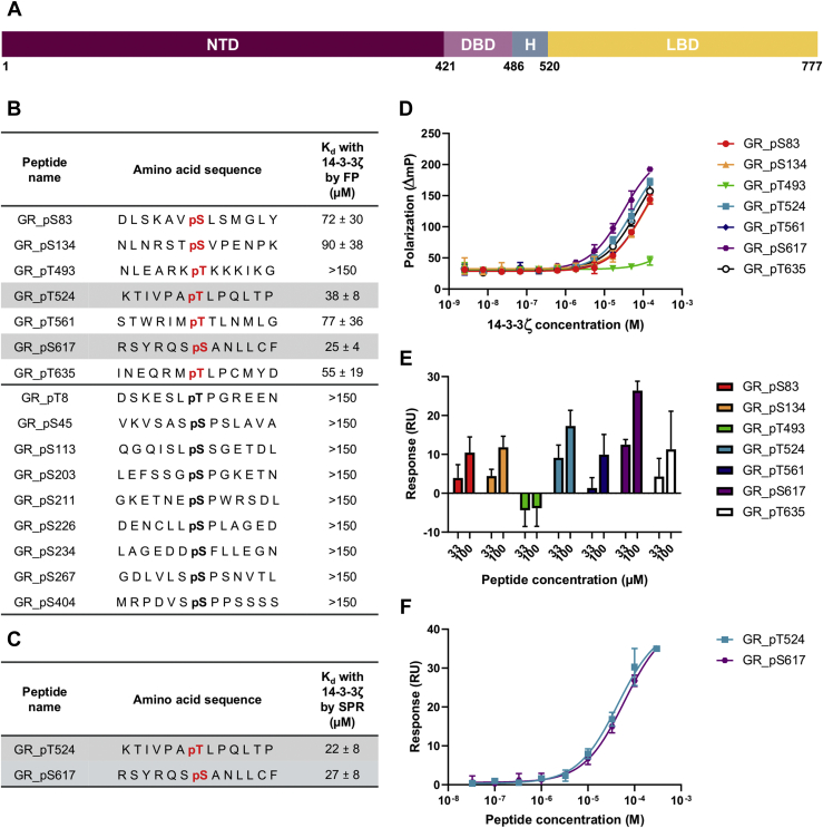
The glucocorticoid receptor (GR) is a ligand-dependent transcription factor that plays a central role in inflammation. The GR activity is also modulated via protein-protein interactions, including binding of 14-3-3 proteins induced by GR phosphorylation. However, the specific phosphorylation sites on the GR that trigger these interactions and their functional consequences are less clear. Hence, we sought to examine this system in more detail. We used phosphorylated GR peptides, biophysical studies, and X-ray crystallography to identify key residues within the ligand-binding domain of the GR, T524 and S617, whose phosphorylation results in binding of the representative 14-3-3 protein 14-3-3ζ. A kinase screen identified misshapen-like kinase 1 (MINK1) as responsible for phosphorylating T524 and Rho-associated protein kinase 1 for phosphorylating S617; cell-based approaches confirmed the importance of both GR phosphosites and MINK1 but not Rho-associated protein kinase 1 alone in inducing GR-14-3-3 binding. Together our results provide molecular-level insight into 14-3-3-mediated regulation of the GR and highlight both MINK1 and the GR-14-3-3 axis as potential targets for future therapeutic intervention.

Keywords: 14-3-3 protein; MINK1 kinase; glucocorticoid receptor; nuclear receptor; phosphorylation; protein–protein interaction.


Conflict of interest statement

Conflict of interest L. D. M., K. E., A. G., A. S., L. W., and M. W. D. P. are employed by and/or own shares in AstraZeneca.

Figures

Figure 1
Figure 1
Interaction of the GR monophosphopeptides with 14-3-3ζ.A, schematic representation of the GR sequence. The number of the residues located at the interface of each GR domain is reported below the sequence. B, amino acid sequences of the monophosphorylated GR peptides centered on the key residues and their binding affinity (Kd) measured by FP. Binding sites of the GR from the literature are depicted in bold black and prediction in bold red. The most potent GR peptides are highlighted in gray. C, Kd of the two most potent monophosphorylated GR peptides measured by SPR. D, concentration–response curves of FP assays of seven peptides centered on predicted 14-3-3 binding sites with 14-3-3ζ. E, affinity of monophosphorylated GR peptides with 14-3-3ζ measured by SPR at two different peptide concentrations (33 and 100 μM). F, concentration–response curves of SPR assays of the two most potent peptides with 14-3-3ζ. All measurements were performed as triplicates, and the error bars represent the SD of these three independent experiments. FP, fluorescence polarization; GR, glucocorticoid receptor; SPR, surface plasmon resonance.
Figure 2
Figure 2
Interaction of the GR doubly phosphorylated peptides with 14-3-3ζ.A, binding affinity (Kd) of the diphosphorylated GR peptides measured by FP. The most potent GR peptide is highlighted in gray. B, binding affinity (Kd) of GR_pT524-pS617 measured by SPR. C, concentration–response curves of FP assays of the four diphosphorylated GR peptides with 14-3-3ζ. D, concentration–response curve of SPR assays of GR_pT524-pS617 with 14-3-3ζ. Measurements were performed as triplicates, and the error bars represent the SD of these three independent experiments. FP, fluorescence polarization; GR, glucocorticoid receptor; SPR, surface plasmon resonance.
Figure 3
Figure 3
Crystal structure of GR_pT524-pS617 bound to 14-3-3ζ dimer.A, the location within the GR sequence of the two 13-mer peptides centered on pT524 and pS617, respectively, and sequence of GR_pT524-pS617. The framed amino acids were assigned in the X-ray structure. B, surface representation of 14-3-3ζ dimer (white and gray solid surface) complexed with GR_pT524-pS617. Residues 520 to 528 are depicted in magenta and residues 614 to 623 in green. The black dashed line shows amino acid residues, not observed in the electron density, connecting the two binding sites. C, details of the interaction between GR_pT524-pS617 and 14-3-3ζ. Polar interactions are depicted as black dotted lines and hydrophobic contracts as magenta or white spheres with a semitransparent surface. GR, glucocorticoid receptor.
Figure 4
Figure 4
Interaction of the GR LBD and full-length GR with pan 14-3-3.A, the location within the GR sequence of pT524 and pS617. B, far-Western blotting overlay of the GR LBD phosphorylated by MINK1 or ROCK1 with BMH1-BMH2-digoxigenin. Unphosphorylated and in vitro phosphorylated GR LBDs were detected using anti-6X His tag antibody. GR LBD-bound 14-3-3 proteins were detected using anti-DIG antibody. C, U2OS cells were transfected with GFP-GR or GFP-GR T524A S617A plasmids and stimulated with calyculin A. Cell lysates were immunoprecipitated with GFP-Trap beads. The GFP-GR and GFP-GR mutant were detected using the anti-GFP antibody, and GR-associated 14-3-3 was detected using anti-pan 14-3-3 antibody. Far-Western blotting overlay was performed by incubation of the GR-containing membrane with BMH1-BMH2-DIG and subsequent detection of GR-bound 14-3-3 protein using the anti-DIG antibody. D, quantification of three independent experiments. E, U2OS cells were transfected with GFP-GR or cotransfected with GFP-GR and FLAG-MINK1 plasmids and treated as mentioned previously. FLAG-MINK1 was detected using the anti-MINK1 antibody. Far-Western blotting overlay was performed as mentioned previously. F, quantification of three independent experiments. Data are normalized to the amount of GFP-GR and GFP-GR mutant from the IP (immunoprecipitation) fraction. The error bars represent the SD of the three independent assays. All p values were obtained using the t test. ∗p < 0.05, ∗∗∗∗p < 0.0001. CaIA, calyculin A; GR, glucocorticoid receptor; LBD, ligand-binding domain; MINK1, misshapen-like kinase 1; OL, overlay; ROCK1, Rho-associated protein kinase 1.

Similar articles

Cited by

References

    1. Kino T. GR-regulating serine/threonine kinases: New physiologic and pathologic implications. Trends Endocrinol. Metab. 2018;29:260–270. - PubMed
    1. Cain D.W., Cidlowski J.A. Immune regulation by glucocorticoids. Nat. Rev. Immunol. 2017;17:233–247. - PMC - PubMed
    1. Souffriau J., Eggermont M., Van Ryckeghem S., Van Looveren K., Van Wyngene L., Van Hamme E., Vuylsteke M., Beyaert R., De Bosscher K., Libert C. A screening assay for selective dimerizing glucocorticoid receptor agonists and modulators (SEDIGRAM) that are effective against acute inflammation. Sci. Rep. 2018;8:12894. - PMC - PubMed
    1. Kuna P., Aurivillius M., Jorup C., Prothon S., Taib Z., Edsbäcker S. Efficacy and tolerability of an inhaled selective glucocorticoid receptor modulator – AZD5423 – in chronic obstructive pulmonary disease patients: Phase II study results. Basic Clin. Pharmacol. Toxicol. 2017;121:279–289. - PubMed
    1. Scheschowitsch K., Leite J.A., Assreuy J. New insights in glucocorticoid receptor signaling-more than just a ligand-binding receptor. Front. Endocrinol. (Lausanne) 2017;8:16. - PMC - PubMed

Publication types

LinkOut - more resources