Review
. 2021 Jan-Jun:296:100553.
doi: 10.1016/j.jbc.2021.100553. Epub 2021 Mar 17.

Beyond the double helix: DNA structural diversity and the PDB

Affiliations
Review

Beyond the double helix: DNA structural diversity and the PDB

Stephen Neidle. J Biol Chem. 2021 Jan-Jun.

Abstract

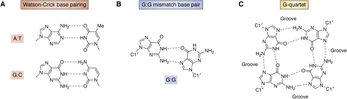
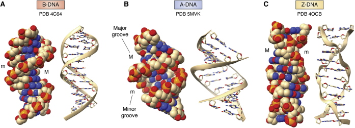
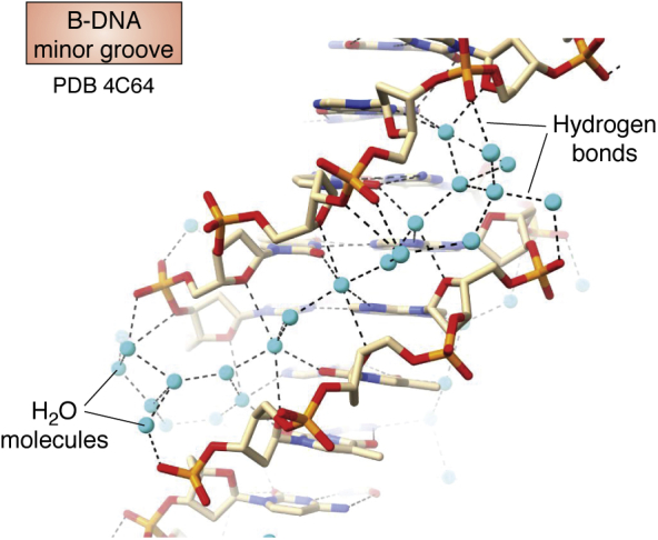
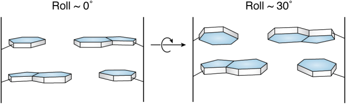
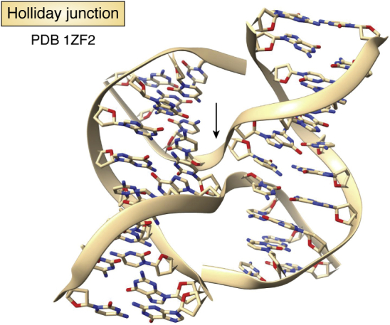
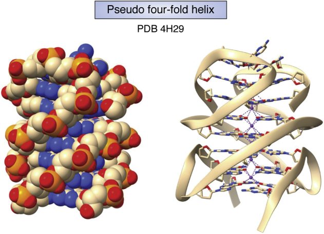
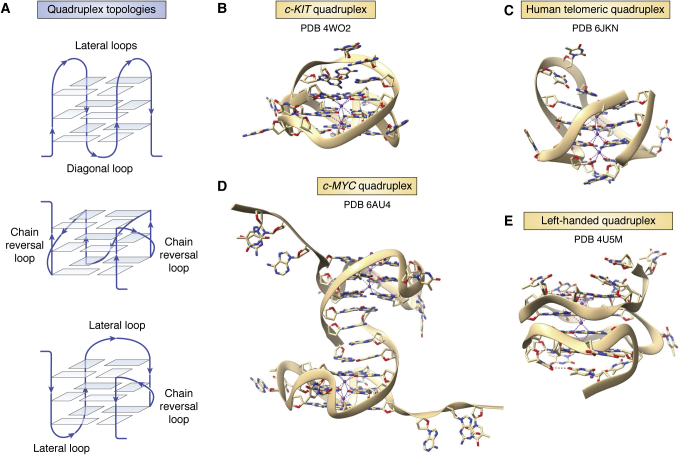
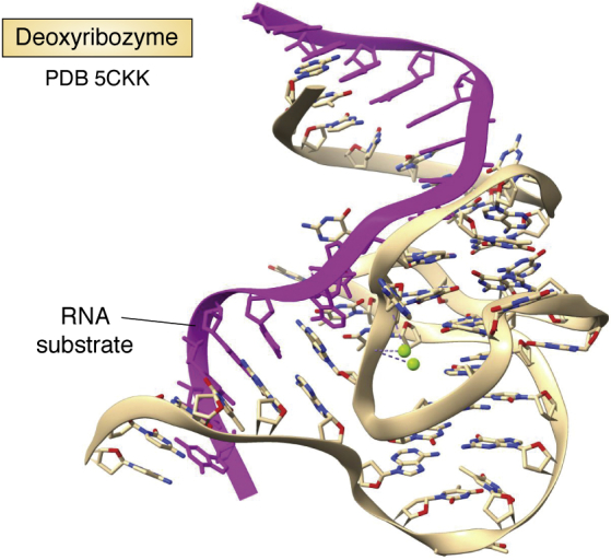
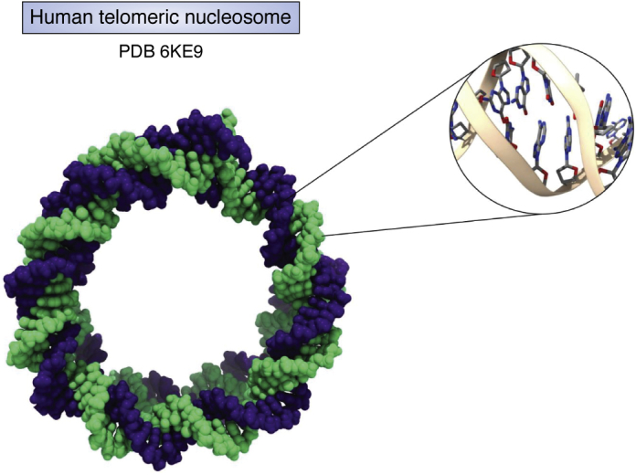
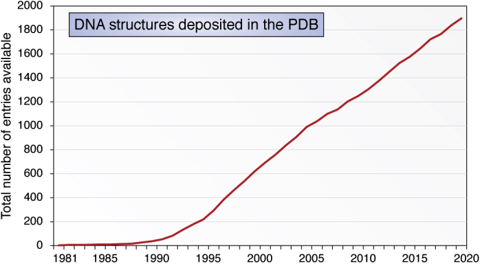
The determination of the double helical structure of DNA in 1953 remains the landmark event in the development of modern biological and biomedical science. This structure has also been the starting point for the determination of some 2000 DNA crystal structures in the subsequent 68 years. Their structural diversity has extended to the demonstration of sequence-dependent local structure in duplex DNA, to DNA bending in short and long sequences and in the DNA wound round the nucleosome, and to left-handed duplex DNAs. Beyond the double helix itself, in circumstances where DNA sequences are or can be induced to unwind from being duplex, a wide variety of topologies and forms can exist. Quadruplex structures, based on four-stranded cores of stacked G-quartets, are prevalent though not randomly distributed in the human and other genomes and can play roles in transcription, translation, and replication. Yet more complex folds can result in DNAs with extended tertiary structures and enzymatic/catalytic activity. The Protein Data Bank is the depository of all these structures, and the resource where structures can be critically examined and validated, as well as compared one with another to facilitate analysis of conformational and base morphology features. This review will briefly survey the major structural classes of DNAs and illustrate their significance, together with some examples of how the use of the Protein Data Bank by for example, data mining, has illuminated DNA structural concepts.

Keywords: DNA; NDB; NMR; PDB; crystal structures; deoxyoligonucleotides; double helix; multistrand helices; quadruplex; sequence-dependent structure.


Conflict of interest statement

Conflict of interest The author declares that he has no conflicts of interest with the contents of this manuscript.

Figures

Figure 1
Figure 1
Various forms of base–base hydrogen bonding observed in DNA crystal structures.A, classic Watson-Crick base pairing as found in unmodified duplex DNAs. B, an example of a base-pair mismatch, as found in some G.G mismatched duplex DNAs. C, the arrangement of eight Hoogsteen hydrogen bonds between the four guanine bases in a G-quartet.
Figure 2
Figure 2
Crystal structures of B, A, and Z form double helices, each showing space-filling and cartoon representations. Major (M) and minor (m) grooves are indicated.A, the structure of the B-DNA Dickerson-Drew dodecamer (18). The two representations are taken from identical viewpoints, with each showing the narrow minor groove in the top part of the helix and the wide major groove in the lower part. These and all subsequent molecular structure figures have used the ChimeraX molecular graphics program (19). B, representations of an A-DNA dodecamer crystal structure (38), showing an approximately 11-fold helix with the characteristic narrow major groove at the center of the view. Base pairs are tilted with respect to the (vertical) helix axis. Although, even though there are local variations in base and base pair morphological parameters, the overall arrangement is close to an A-DNA helix derived from fiber diffraction. C, representations of the crystal structure of the left-handed Z-DNA sequence d(CGCGCGCGCGCG) (48). Note the characteristic irregular zig-zag feature of the phosphodiester backbone shown in ribbon cartoon form. PDB, Protein Data Bank.
Figure 3
Figure 3
Detail of the water structure in the B-DNA dodecamer minor groove (18), showing the water molecules (in cyan) and hydrogen bonds to waters and DNA. PDB, Protein Data Bank.
Figure 4
Figure 4
A schematic representation of roll between two successive base pairs. LHS: the roll is ca 0°. RHS: the roll, of ca 30°, is opening the base pair toward the minor groove, i.e., toward the viewer. The cross-over point is indicated by an arrow.
Figure 5
Figure 5
Structure of a Holliday junction (43), drawn in cartoon form, with the strand cross-over point in the center indicated by the arrow. The two B-DNA helices are inclined at an angle of ca 40°. PDB, Protein Data Bank.
Figure 6
Figure 6
van der Waals and cartoon depictions of a four-stranded right-handed quadruplex pseudo helix, as observed in the central core of seven stacked G-quartets in the crystal structure of the BRAF promoter quadruplex (77). All external looped-out adenine bases have been removed to enhance clarity. This bimolecular quadruplex is formed by two antiparallel strands, analogous to a duplex; however, these are highly G-rich and fold back to place the Gs in register and form the successive G-quartets and a four-stranded arrangement with four grooves. PDB, Protein Data Bank. Coordinated potassium ions are visible in the centre of the G-helix.
Figure 7
Figure 7
Quadruplex folds.A, Schematic figures of three distinct quadruplex topologies, showing different loop types. The backbones in each case are colored blue, with strand directions shown by arrows. Human telomeric quadruplexes are polymorphic in solution and can adopt these and several other topologies (59–61). B, cartoon representation of the quadruplex formed from a promoter sequence in the c-KIT gene (76). Note the large A:G base-paired loop at the top of the structure. C, cartoon representation of the antiparallel chair arrangement formed by a human telomeric quadruplex (65), with three lateral loops. D, cartoon representation of the crystal structure of the c-MYC quadruplex (78). The two independent molecules in the crystallographic asymmetric unit are shown. Both have all-parallel topology, as in the human telomeric quadruplex (66), but with distinctive loops suitable for selective ligand binding. E, cartoon representation of a left-handed quadruplex (81). Note the narrow zig-zag groove parallel to the G-quartets. PDB, Protein Data Bank.
Figure 8
Figure 8
Ribbon cartoon representation of the crystal structure of a deoxyribozyme (91). Its RNA substrate sequence is colored purple. PDB, Protein Data Bank.
Figure 9
Figure 9
The structure of the DNA component of a human telomeric nucleosome (105), shown in space-filling representation with the two strands in separate colors, together with a magnified view of a typical region of DNA curvature, showing changes from ideality in base pair morphology such as roll, tilt, and twist.
Figure 10
Figure 10
Year-on-year increases in the number of DNA structures deposited in the PDB, in the period 1981 to 2020. I am grateful to Dr Cathy Lawson, the current Director of the NDB, for this information. NDB, Nucleic Acid Database; PDB, Protein Data Bank.

References

    1. Neidle S. Academic Press; London: 2008. Principles of Nucleic Acid Structure.
    1. Watson J.D., Crick F.H. Molecular structure of nucleic acids; a structure for deoxyribose nucleic acid. Nature. 1953;171:737–738. - PubMed
    1. Wilkins M.H., Stokes A.R., Wilson H.R. Molecular structure of deoxypentose nucleic acids. Nature. 1953;171:738–740. - PubMed
    1. Franklin R.E., Gosling R.G. Molecular configuration in sodium thymonucleate. Nature. 1953;171:740–741. - PubMed
    1. Arnott S., Hutchinson F., Spencer M., Wilkins M.H., Fuller W., Langridge R. X-ray diffraction studies of double helical ribonucleic acid. Nature. 1966;211:227–232. - PubMed

LinkOut - more resources