. 2021 Jul 7;29(7):2281-2293.
doi: 10.1016/j.ymthe.2021.03.014. Epub 2021 Mar 17.

Targeting of miR-33 ameliorates phenotypes linked to age-related macular degeneration

Affiliations

Targeting of miR-33 ameliorates phenotypes linked to age-related macular degeneration

Gopalan Gnanaguru et al. Mol Ther. .

Abstract

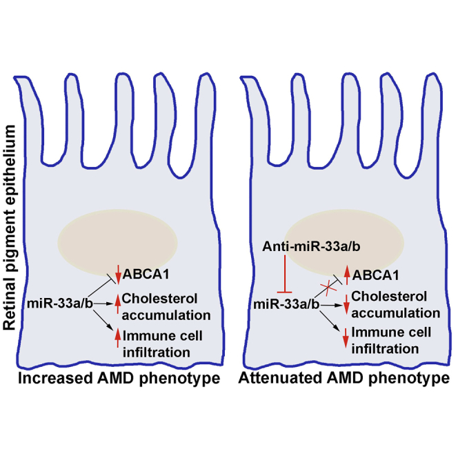
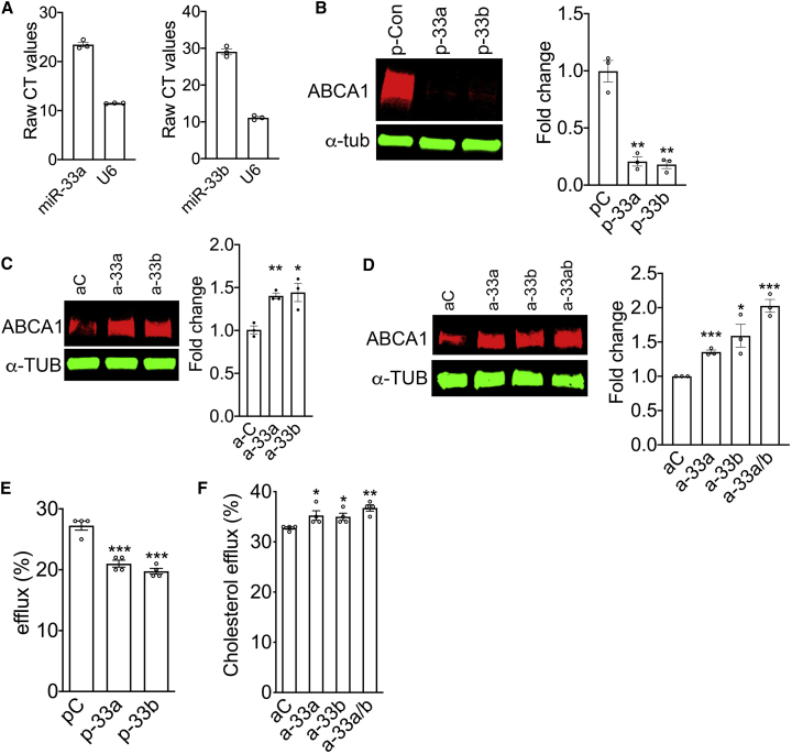
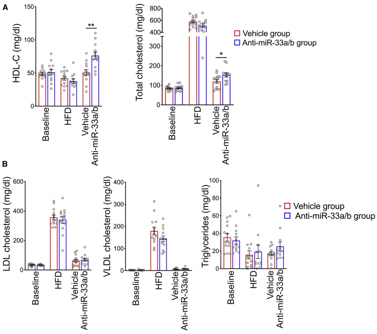
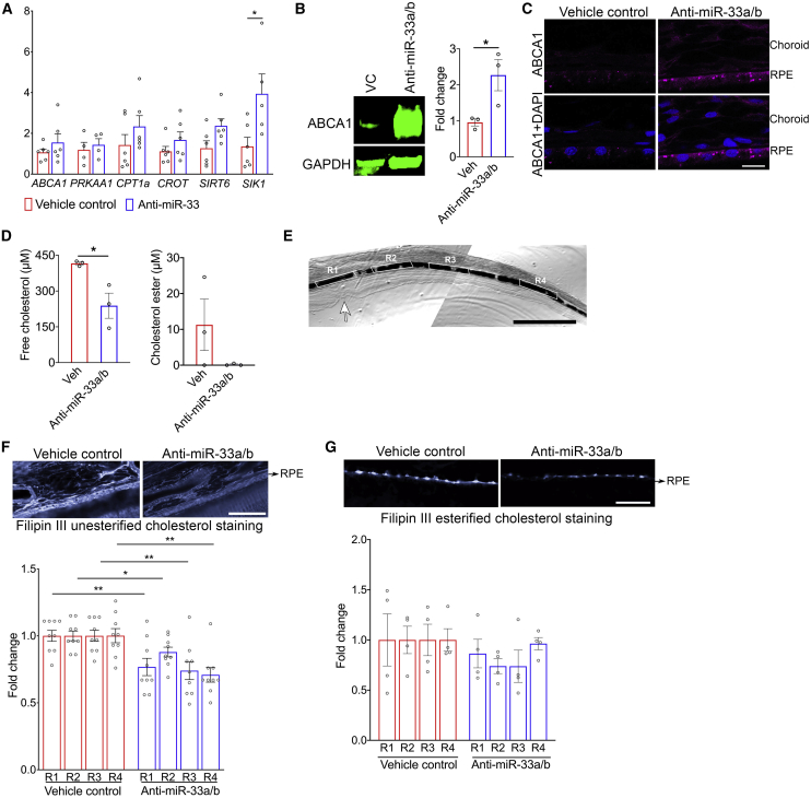
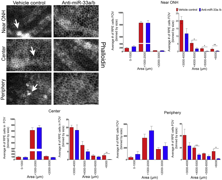
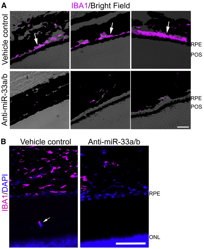
Abnormal cholesterol/lipid homeostasis is linked to neurodegenerative conditions such as age-related macular degeneration (AMD), which is a leading cause of blindness in the elderly. The most prevalent form, termed "dry" AMD, is characterized by pathological cholesterol accumulation beneath the retinal pigment epithelial (RPE) cell layer and inflammation-linked degeneration in the retina. We show here that the cholesterol-regulating microRNA miR-33 was elevated in the RPE of aging mice. Expression of the miR-33 target ATP-binding cassette transporter (ABCA1), a cholesterol efflux pump genetically linked to AMD, declined reciprocally in the RPE with age. In accord, miR-33 modulated ABCA1 expression and cholesterol efflux in human RPE cells. Subcutaneous delivery of miR-33 antisense oligonucleotides (ASO) to aging mice and non-human primates fed a Western-type high fat/cholesterol diet resulted in increased ABCA1 expression, decreased cholesterol accumulation, and reduced immune cell infiltration in the RPE cell layer, accompanied by decreased pathological changes to RPE morphology. These findings suggest that miR-33 targeting may decrease cholesterol deposition and ameliorate AMD initiation and progression.

Keywords: ABCA1; age-related macular degeneration; cholesterol; geographic atrophy; inflammation; microRNA; retinal pigment epithelial cells.


Conflict of interest statement

Declaration of interests The authors declare no competing interests.

Figures

None
Graphical abstract
Figure 1
Figure 1
miR-33 modulated ABCA1 expression and cholesterol efflux in RPE cells (A) The expression of miR-33a and miR-33b was analyzed by quantitative RT-PCR in primary human RPE cells (n = 3). (B) Western blot showing the expression of ABCA1 in primary human RPE cells 72 h after transfection with precursor miR control, miR-33a, or miR-33b (n = 3). (C) Western blotting demonstrating ABCA1 in primary human RPE cells 72 h post-transfection with control, anti-miR-33a, or anti-miR-33b (n = 3). (D) Western blotting demonstrating ABCA1 in ARPE-19 cells 72 h post-transfection with control, anti-miR-33a, anti-miR-33b, or anti-miR33a/b (n = 3). (E) TopFluor cholesterol efflux was measured in ARPE-19 cells transfected with precursor control miR, miR-33a, or miR-33b. (F) TopFluor cholesterol efflux was assessed in ARPE-19 cells ∼60 h after transfection with scrambled control, anti-miR-33a, anti-miR-33b, or-miR-33a/b ASO. pC, precursor scrambled control miR; aC, anti-miR control. All error bars represent ±SEM. (B–D) Expression levels were normalized to α-tubulin loading control, and statistical significance between groups was calculated by unpaired t test. (E and F) Each experiment was performed in quadruplicate and repeated ≥3 times, and statistical significance between groups was calculated by unpaired t test. ∗p < 0.05, ∗∗p < 0.01, ∗∗∗p < 0.001.
Figure 2
Figure 2
Anti-miR-33a/b ASO treatment improves plasma HDL-C and total cholesterol in NHPs (A) Plasma HDL-C and total cholesterol were measured in NHPs fed a high-fat/cholesterol diet for 20 months and then switched to a regular chow diet and injected with anti-miR-33 ASO or vehicle for 6 weeks (n = 12/group). (B) Serum AST, ALT, bilirubin, and uric acid levels were measured to monitor liver and kidney functions in mice that were fed a high-fat/cholesterol diet and injected with scrambled control or anti-miR-33 LNA ASO (n = 12/group). All error bars represent ±SEM. Statistical differences between vehicle control and anti-miR-33a/b ASO-injected mice were calculated by unpaired t test. ∗p < 0.05 and ∗∗p < 0.01.
Figure 3
Figure 3
Anti-miR-33a/b ASO treatment increased miR-33 target gene expression levels and ABCA1 protein localization in NHP RPE cell layer (A) Expression levels of ABCA1, PRKAA1, CPT1A, CROT, SIRT6, and SIK1 were measured by quantitative RT-PCR in RPE cells isolated from NHPs injected with anti-miR-33 ASO or vehicle for 6 weeks (n ≤ 4). mRNA expression levels were normalized to PPIH or HPRT1. (B) Protein blot showing the ABCA1 protein expression in RPE/choroid lysate extracted from vehicle or anti-miR-33 injected NHP (n = 3) and the bar graph showing the relative fold change. (C) Retinal cryosections prepared from NHPs that were treated with vehicle or anti-miR-33 ASO by subcutaneous injections were immunostained for ABCA1 and DAPI nuclear stain (n = 3). Scale bar: 10 μm. (D) Free and esterified cholesterol levels were measured in the lipid extracts from RPE/choroid of NHPs that were injected with anti-miR-33 ASO or vehicle for 6 weeks (n = 3). (E) NHP retinal sections showing four highlighted regions (R1–4) from the fovea to the periphery were chosen to quantify filipin III staining in the RPE cell layer of vehicle- or anti-miR-33 ASO-treated NHP retinal sections. Arrow points to fovea; scale bar: 1 mm. (F) Retinal sections of NHPs that were injected with anti-miR-33a/b ASO or vehicle for 6 weeks were stained with filipin III to label unesterified cholesterol. Four regions (R1–4) from the fovea to the periphery shown in (E) chosen to quantify filipin III staining in the RPE cell layer of vehicle- or anti-miR-33 ASO-treated NHP retinal sections (n = 9). Scale bar: 50 μm. (G) Retinal sections of NHPs that were injected with anti-miR-33 ASO or vehicle for 6 weeks were pretreated with cholesterol esterase and then stained with filipin III to label esterified cholesterol. Four regions (R1–4) from the fovea to the periphery shown in (E) chosen to quantify filipin III staining in the RPE cell layer of vehicle- or anti-miR-33 ASO-treated NHP retinal sections (n = 4). Scale bar: 50 μm. All error bars represent ± SEM. Statistical differences between vehicle- and anti-miR-33 ASO-injected NHPs were calculated by unpaired t test. ∗p < 0.05, ∗∗p < 0.01, ∗∗∗p < 0.001.
Figure 4
Figure 4
Anti-miR-33a/b ASO treatment reduced abnormal RPE cytoskeletal organization in the RPE cell layer of NHPs fed a high-fat/cholesterol diet RPE flatmounts prepared from NHPs that received subcutaneous injections of vehicle or anti-miR-33 ASO for 6 weeks were stained with phalloidin and examined for RPE cytoskeletal organization, and then RPE cell size was quantified and segmented in the area closer to the optic nerve head (ONH), center, and periphery. Arrows in the top panel indicate enlarged RPE cells (n ≤ 7). Scale bar: 100 μm. RPE cell size was quantified in field of view (FOV) and represented in the bar graphs. All error bars represent ±SEM. Statistical differences between vehicle- and anti-miR-33 ASO-injected NHPs were calculated by unpaired t test. ∗p < 0.05, ∗∗p < 0.01, ∗∗∗p < 0.001.
Figure 5
Figure 5
Anti-miR-33a/b ASO treatment reduced immune cell infiltration in RPE-photoreceptor and RPE layers (A) IBA1 staining (magenta) and superimposed brightfield (BF) revealing IBA1 positive cells in the sub-RPE layers in vehicle-treated NHP retinal sections, while low IBA1-positive staining is seen in the sub-RPE-choroid layer of anti-miR-33 ASO-injected NHP retinal sections. Scale bar: 20 μm. POS, photoreceptor outer segment. (B) Representative IBA1 (magenta) and superimposed DAPI (blue) staining revealing IBA1-positive cell in the RPE-photoreceptor layer in vehicle-treated NHP retinal sections (white arrow), while no IBA1-positive staining is seen in the RPE-photoreceptor layer of anti-miR-33 ASO-injected NHP retinal sections. Scale bar: 20 μm. ONL, outer nuclear layer of photoreceptor cells.

Similar articles

Cited by

References

    1. Xu Q., Cao S., Rajapakse S., Matsubara J.A. Understanding AMD by analogy: systematic review of lipid-related common pathogenic mechanisms in AMD, AD, AS and GN. Lipids Health Dis. 2018;17:3. - PMC - PubMed
    1. Curcio C.A., Johnson M., Huang J.D., Rudolf M. Apolipoprotein B-containing lipoproteins in retinal aging and age-related macular degeneration. J. Lipid Res. 2010;51:451–467. - PMC - PubMed
    1. Curcio C.A., Johnson M., Rudolf M., Huang J.D. The oil spill in ageing Bruch membrane. Br. J. Ophthalmol. 2011;95:1638–1645. - PMC - PubMed
    1. Moreira E.F., Larrayoz I.M., Lee J.W., Rodríguez I.R. 7-Ketocholesterol is present in lipid deposits in the primate retina: potential implication in the induction of VEGF and CNV formation. Invest. Ophthalmol. Vis. Sci. 2009;50:523–532. - PMC - PubMed
    1. McKay G.J., Patterson C.C., Chakravarthy U., Dasari S., Klaver C.C., Vingerling J.R., Ho L., de Jong P.T., Fletcher A.E., Young I.S. Evidence of association of APOE with age-related macular degeneration: a pooled analysis of 15 studies. Hum. Mutat. 2011;32:1407–1416. - PMC - PubMed

Publication types