. 2021 Mar 19;13(7):9874-9899.
doi: 10.18632/aging.202740. Epub 2021 Mar 19.

DNASE1L3 arrests tumor angiogenesis by impairing the senescence-associated secretory phenotype in response to stress

Affiliations

DNASE1L3 arrests tumor angiogenesis by impairing the senescence-associated secretory phenotype in response to stress

Deliang Guo et al. Aging (Albany NY). .

Abstract

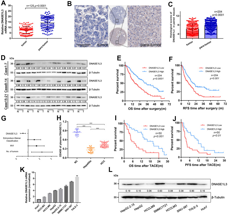
Hepatocellular carcinoma (HCC) is one of the most challenging and aggressive cancers with limited treatment options because of tumor heterogeneity. Tumor angiogenesis is a hallmark of HCC and is necessary for tumor growth and progression. DNA damage stress and its associated deoxyribonuclease1-like 3 (DNASE1L3) are involved in HCC progression. Here, we explored the influence mechanism of DNASE1L3 on tumor angiogenesis under DNA damage stress in vitro and in vivo. DNASE1L3 was found downregulated and negatively correlated with poor prognosis of resectable and unresectable HCC patients. The tissue microarray of HCC revealed the negative association between DNASE1L3 and cancer vasculature invasion. Mechanistically, DNASE1L3 was found to relieve cytoplasmic DNA accumulation under DNA damage stress in HCC cell lines, in turn cell senescence and senescence-associated secretory phenotype were arrested via the p53 and NF-κB signal pathway, and hence, tumor angiogenesis was impaired. Furthermore, we found that DNASE1L3 excised these functions by translocating to the nucleus and interacting with H2BE under DNA damage stress using co-immunoprecipitation and fluorescence resonance energy transfer assay. In conclusion, DNASE1L3 inhibits tumor angiogenesis via impairing the senescence-associated secretory phenotype in response to DNA damage stress.

Keywords: DNASE1L3; angiogenesis; hepatocellular carcinoma; senescence.


Conflict of interest statement

CONFLICTS OF INTEREST: All the authors declared no conflicts of interest.

Figures

Figure 1
Figure 1
Downregulated DNASE1L3 is positively correlated with prognosis of resectable or unresectable HCC. (A) The transcriptional level of DNASE1L3 was down-regulated in HCC tissues(n=125) compared to paired para-tumor tissues as tested by RT-qPCR. (B) Representative images of IHC staining for DNASE1L3 in HCC and adjacent normal tissues (scale bar, 50 μm). (C) The histological scores of DNASE1L3 in 204 paired tissues of HCC was evaluated. (D) The translational level of DNASE1L3 between HCC tissues and paired adjacent non-tumor tissues from 21 patients were identified by western blotting. (“T” for tumor, “N” for non-tumor). (E, F) Kaplan-Meier survival curves of OS and RFS time for 204 patients with HCC. (G) Forest plot of risk factors of the OS time using multivariate Cox regression analysis. (H) Comparison of plasma levels of DNASE1L3 between patients of HCC(n=50), patients of hepatitis only(n=27) and healthy individuals(n=18). (I, J) Kaplan-Meier survival curves of OS and PFS time for 50 patients with inoperable HCC. (K) RT-qPCR analysis of DNASE1L3 in HCC cell lines and a normal liver cell line (THLE-3). (L) Western blot analysis of DNASE1L3 protein expression in HCC cell lines and a normal liver cell line (THLE-3).
Figure 2
Figure 2
Overexpression of DNASE1L3 relieves cytoplasmic DNA accumulation under DDR activation. (A) Cytoplasmic accumulation of nuclear DNA in differentially treated cells were assessed, representative images were shown (green, γH2AX; blue, DAPI; Scale bars, 10 μm). (B) Quantification of DNA damage foci (DDF). The number of DDF per cell falls into each of the 0, 1-3, 4-10, and >10 counting categories. At least 100 cells counted per group. (C, D) qPCR analysis of chromosomal DNA in cytoplasmic fraction of cells treated in different group.
Figure 3
Figure 3
Overexpression of DNASE1L3 relieves cell senescence and SASP under DDR activation. (A) Representative images of SA-β-Gal staining of cells in differently treated groups (scale bar, 200 μm). (B) Statistics of SA-β-Gal staining cells in differently treated groups. (C, D) Remnant cell number were analyzed by cell counts enumerated from differently treated groups at various time points. (E) Transcriptional level of canonical SASP factors in differently treated groups, with values normalized to the vector control per factor. The heatmap for the mean RT-qPCR data is shown. (F) Immunoblot analysis of inducible expression change of senescence associated signal pathway and downstream proteins including p53, p65, SPINK1 and AREG in different treated groups. (G) Dual luciferase assay showed activated NF-kB signaling in different treated groups. (H, I) ELISA analysis of IL-6, IL-8 secretion in supernatants from cells in different treated groups.
Figure 4
Figure 4
DNASE1L3 impairs angiogenesis through the differential expression of SASP under DDR activation. (A) Tissue microarray was stained with anti-DNASE1L3 and anti-CD34 antibody, low expression of DNASE1L3 was considerably associated with cancer vasculature invasion. (scale bar, 100 μm). (B, C) Tumor tissues of DNASE1L3 intermediately or highly expressed were observed to be linked with lower vasculature invasion. (scale bar, 100 μm). (D) The histological scores of DNASE1L3 and the CD34 Chalkley counts in HCC tissues was analyzed and showed a significant negative association. (E, F) The motility of HUVECs were assessed by wound healing assay, the supernatants from cells in differently treated groups were added into the culture of HUVECs, images were taken at 0h and 24h (scale bar, 100 μm). (G, H) The cellular migration ability of HUVECs were determined by the transwell migration assay. The supernatants from cells in differently treated groups were added into the lower chamber, images were taken after 24h of incubation (scale bar, 100 μm). (I, J) The tube formation ability of HUVECs were determined by tube formation assay. The supernatants from cells in differently treated groups were added into the culture, images were taken after 6h of incubation (scale bar, 100 μm). The results show the means ± SD from at least three separate experiments.
Figure 5
Figure 5
DNASE1L3 interacts with H2BE. (A, B) Immunoblot analysis of DNASE1L3 in the subcellular localization under DNA damage using nucleocytoplasmic separation in two cell lines. (C) The result of blotting confirmed that DNASE1L3 translocated to the nucleus in response to DDR activation. (D) Lysates of 293T cells overexpressing Flag-H2BE and/or cMyc-DNASE1L3 were subjected to reciprocal co-immunoprecipitation (co-IP) to detect protein interaction. (E) HepG2 and HCCLM3 cell lysates were subjected to co-IP and immunoblot to detect endogenous H2BE and DNASE1L3 interaction. (F) FRET assay for DNASE1L3-H2BE interactions in living cells. FRET efficiency were calculated by Leica TCS SP8 software (FRETeff= (Dpost-Dpre)/Dpost). White circles identify FRET area. (G) Co-IP of cMyc-DNASE1L3 and Flag-tagged H2BE or H2BEΔ with N-terminal region (amino acids 1-28) deleted.
Figure 6
Figure 6
DNASE1L3 impairs angiogenesis by interacting with H2BE. (A) qPCR analysis of chromosomal DNA in the cytoplasmic fraction of differently treated HepG2 cells. (B) Representative images of SA-β-Gal staining of cells in differently treated groups (scale bar, 100 μm). (C) Statistics of SA-β-Gal staining cells in differently treated groups. (D) Immunoblot analysis of inducible expression change of senescence associated signal pathway and downstream proteins including p53, p65, SPINK1 and AREG in differently treated groups. (E, F) The motility of HUVECs were assessed by wound healing assay, the supernatants from cells in different treated groups were added into the culture of HUVECs, images were taken at 0h and 24h (scale bar, 100 μm). (G, H) The cellular migration ability of HUVECs were determined by the transwell migration assay. Cell supernatants from in differently treated groups were added into the lower chamber, images were taken after 24h of incubation (scale bar, 100 μm). (I, J) The tube formation ability of HUVECs were determined by tube formation assay. Cell supernatants from differently treated groups were added into the culture, images were taken after 6h of incubation (scale bar, 100 μm). The results show the means ± SD from at least three separate experiments.
Figure 7
Figure 7
DNASE1L3 arrests tumor angiogenesis by regulating the senescence-associated secretory phenotype in response to stress. (A) Matrigel plugs supplemented with cell supernatants from differently treated groups were subcutaneously injected into mice. (B) Hemoglobin content was assessed at day 10 after injection. (C) Representative images of CD34 staining of the matrigel plugs in differently treated groups (scale bar, 200 μm). Gross view observation of the matrigel plugs were shown in the bottom right corner of each figure. (D) CD34 positive area was evaluated in each group. (E) Constructed subcutaneous tumor model was used to investigate the role of DNASE1L3 in tumor angiogenesis. (F) Vascular density was quantified by CD34 Chalkley count under IHC. (G) Representative images of CD34 staining of subcutaneous tumors in differently treated groups (scale bar, 200 μm).
Figure 8
Figure 8
Model depicting DNASE1L3 regulation of tumor angiogenesis by controlling the senescence-associated secretory phenotype in response to stress.

Similar articles

Cited by

References

    1. Bray F, Ferlay J, Soerjomataram I, Siegel RL, Torre LA, Jemal A. Global cancer statistics 2018: GLOBOCAN estimates of incidence and mortality worldwide for 36 cancers in 185 countries. CA Cancer J Clin. 2018; 68:394–424. 10.3322/caac.21492 - DOI - PubMed
    1. Galle PR, Tovoli F, Foerster F, Wörns MA, Cucchetti A, Bolondi L. The treatment of intermediate stage tumours beyond TACE: from surgery to systemic therapy. J Hepatol. 2017; 67:173–83. 10.1016/j.jhep.2017.03.007 - DOI - PubMed
    1. Reig M, da Fonseca LG, Faivre S. New trials and results in systemic treatment of HCC. J Hepatol. 2018; 69:525–33. 10.1016/j.jhep.2018.03.028 - DOI - PubMed
    1. Lyu N, Kong Y, Mu L, Lin Y, Li J, Liu Y, Zhang Z, Zheng L, Deng H, Li S, Xie Q, Guo R, Shi M, et al.. Hepatic arterial infusion of oxaliplatin plus fluorouracil/leucovorin vs. sorafenib for advanced hepatocellular carcinoma. J Hepatol. 2018; 69:60–69. 10.1016/j.jhep.2018.02.008 - DOI - PubMed
    1. Abou-Alfa GK, Qin S, Ryoo BY, Lu SN, Yen CJ, Feng YH, Lim HY, Izzo F, Colombo M, Sarker D, Bolondi L, Vaccaro G, Harris WP, et al.. Phase III randomized study of second line ADI-PEG 20 plus best supportive care versus placebo plus best supportive care in patients with advanced hepatocellular carcinoma. Ann Oncol. 2018; 29:1402–08. 10.1093/annonc/mdy101 - DOI - PubMed

Publication types

MeSH terms

Substances