. 2021 Mar 19;13(7):9646-9664.
doi: 10.18632/aging.202708. Epub 2021 Mar 19.

BMP5 silencing inhibits chondrocyte senescence and apoptosis as well as osteoarthritis progression in mice

Affiliations

BMP5 silencing inhibits chondrocyte senescence and apoptosis as well as osteoarthritis progression in mice

Yan Shao et al. Aging (Albany NY). .

Abstract

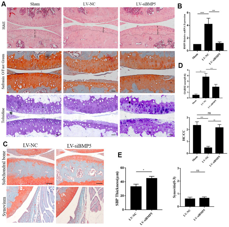
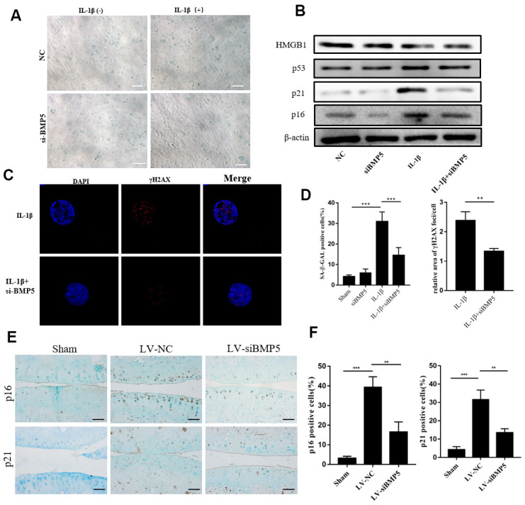
In this study, we using the in vivo destabilization of the medial meniscus (DMM) mouse model to investigate the role of bone morphogenetic protein 5 (BMP5) in osteoarthritis (OA) progression mediated via chondrocyte senescence and apoptosis. BMP5 expression was significantly higher in knee articular cartilage tissues of OA patients and DMM model mice than the corresponding controls. The Osteoarthritis Research Society International scores based on histological staining of knee articular cartilage sections were lower in DMM mice where BMP5 was knocked down in chondrocytes than the corresponding controls 4 weeks after DMM surgery. DMM mice with BMP5-deficient chondrocytes showed reduced levels of matrix-degrading enzymes such as MMP13 and ADAMTS5 as well as reduced cartilage destruction. BMP5 knockdown also decreased chondrocyte apoptosis and senescence by suppressing the activation of p38 and ERK MAP kinases. These findings demonstrate that BMP5 silencing inhibits chondrocyte senescence and apoptosis as well as OA progression by downregulating activity in the p38/ERK signaling pathway.

Keywords: BMP5; chondrocyte; osteoarthritis; p38/ERK; senescence.


Conflict of interest statement

CONFLICTS OF INTEREST: The authors declare that they have no conflicts of interest.

Figures

Figure 1
Figure 1
Bmp5 is overexpressed in knee articular cartilage tissues of OA patients. (A) Representative images show Safranin O-Fast Green staining and BMP5 immunohistochemical staining in the knee articular cartilage tissues from healthy subjects and OA patients (n=6 each). Scale bar: 10 μm. (B) OARSI scores based on the histochemical staining and corresponding BMP5 immunohistochemical staining of human knee articular cartilage tissue sections from healthy and osteoarthritis patients. (C) Representative western blots show BMP5 protein expression in the knee articular cartilage samples from healthy individuals and OA patients. (D) Representative images show Safranin O-Fast Green staining and BMP5 immunohistochemical staining in the knee articular cartilage sections from sham, DMM-2W and DMM-4W model mice (n=5 each). Scale bars: 10 μm (first line) and 5 μm (others). (E) Representative western blots show the levels of BMP5 protein in the knee articular cartilage sections from sham, DMM-2W and DMM-4W model mice. (F) ELISA assay results show joint fluid BMP5 levels in the healthy and OA patients. (G) BMP5 immunohistochemical staining scores and OARSI scores of the DMM-2W, DMM-4W, and sham model mice are shown. All data are represented as means ± SD.
Figure 2
Figure 2
Deletion of BMP5 attenuates osteoarthritis in the in vivo DMM model mice. (A) Representative images show hematoxylin and eosin (H&E) staining, toluidine staining and Safranin O-Fast Green staining of knee cartilage tissue sections from LV-siBMP5 and LV-siNC group mice (n=5 per group). (B) RT-PCR results show BMP5 mRNA levels in LV-siBMP5 and LV-siNC group mice at 2 weeks post-DMM surgery (n = 5 per group). (C) Representative images show synovitis and SBP thickness in the sham-operated, DMM plus LV-siNC, and DMM plus LV-siBMP5 groups of mice at 4 weeks post-DMM operation. (D) OARSI scores and hyaline cartilage/calcified cartilage ratios in sham-operated, DMM plus LV-siNC, and DMM plus LV-siBMP5 groups of mice at 4 weeks post-DMM operation. (E) Quantitative analysis shows the status of synovitis and SBP thickness in the sham-operated, DMM plus LV-siNC, and DMM plus LV-siBMP5 groups of mice at 4 weeks post-DMM operation. All data are represented as the means ± SD (n=5 per group); scale bars: 5 μm.
Figure 3
Figure 3
BMP5 regulates in vivo and in vitro chondrocyte catabolism. (A) QRT-PCR assay results show mRNA expression levels of pro-inflammatory cytokines (TNF-α, IL-1β), anabolic factors (type II collagen and SOX9), and catabolic factors (MMP13 and ADAMTS-5) in control and BMP-knockdown murine chondrocytes, treated with IL-1β or not. (B) Representative western blots show expression levels of MMP13, Runx2, and Col2 proteins in control and BMP-knockdown murine chondrocytes. (C) Representative immunohistochemical images and (D) quantitative analyses of the IHC results show MMP13, Col2a, and TIMP2 expression in the knee articular cartilage sections from sham-operated, DMM plus LV-siNC, and DMM plus LV-siBMP5 groups of mice at 4 weeks post-DMM operation. All data are represented as means ± SD (n=5 per group).
Figure 4
Figure 4
BMP5 knockdown inhibits in vitro and in vivo chondrocyte senescence in the in vitro and in vivo OA models. (A) The SA-β gal staining assay results in IL-1β-treated control and BMP5 knockdown murine chondrocytes. (B) Representative western blots show the levels of HMGB1, p53, p16, and p21 proteins in IL-1β-treated control and BMP5 knockdown murine chondrocytes. (C) Representative immunofluorescence images show γH2AX (red) staining in control and BMP5 knockdown murine chondrocytes treated with IL-1β. The chondrocytes were counterstained with DAPI (blue), a DNA binding dye. (D) Quantitative analyses show SA-β gal staining assay and γH2AX (red) staining in treated as above. (E, F) Representative immunohistochemical staining and quantitative analysis of p16 and p21 expression in the knee articular cartilage tissues from sham-operated, DMM plus LV-siNC, and DMM plus LV-siBMP5 groups of mice at 4 weeks post-DMM operation. All data are represented as the means ± SD (n=5); scale bars: 5 μm; **P<0.01; ***P<0.001.
Figure 5
Figure 5
BMP5 silencing protects against chondrocyte apoptosis in both in vivo and in vitro OA models. (A) Representative images show TUNEL staining assay in control and BMP5 knockdown murine chondrocytes treated with IL-1β. Scale bar: 5μm. (B) Quantitative analysis shows the total numbers of TUNEL-positive control and BMP5 knockdown chondrocytes treated with IL-1β. (C) Representative western blot images show BCL2 and cleaved-caspase3 protein levels in control and BMP5 knockdown chondrocytes treated with IL-1β. (D) Representative images and (E) quantitative analyses show TUNEL staining and BCl-2 immunohistochemical staining results in the knee articular cartilage tissues from sham-operated, DMM plus LV-siNC, and DMM plus LV-siBMP5 groups of mice at 4 weeks post-DMM operation. Scale bar: 5μm. All data are represented as the means ± SD (n=5); Scale bars: 5μm.
Figure 6
Figure 6
BMP5 regulates osteoarthritis progression via p38/ERK signaling pathway. (A) Representative western blots show the expression levels of total ERK, p-ERK, total p38, and p-38 proteins in control and BMP5 knockdown chondrocytes mouse chondrocytes treated with IL-1β. (B, D) Representative western blots and quantification show the expression levels of total ERK, p-ERK, total p38, and p-p38 in mouse chondrocytes only treated with different concentrations of rhBMP5(50,100,300ng/ml). (C) Western blot analysis shows the levels of total ERK, p-ERK, total p38, p-p38, p16, cleaved-caspase3, and MMP13 proteins in mouse chondrocytes treated with 100ng/ml rhBMP5, 10 μM PD98059 (MAPK inhibitor) or co-treatment with rhBMP5 and 10 μM PD98059. (E) A schematic working model shows the potential role of Bmp5 in regulating senescence and apoptosis of chondrocytes.

Similar articles

Cited by

References

    1. Hunter DJ, Schofield D, Callander E. The individual and socioeconomic impact of osteoarthritis. Nat Rev Rheumatol. 2014; 10:437–41. 10.1038/nrrheum.2014.44 - DOI - PubMed
    1. Hunter DJ, Bierma-Zeinstra S. Osteoarthritis. Lancet. 2019; 393:1745–59. 10.1016/S0140-6736(19)30417-9 - DOI - PubMed
    1. Roseti L, Desando G, Cavallo C, Petretta M, Grigolo B. Articular Cartilage Regeneration in Osteoarthritis. Cells. 2019; 8:1305. 10.3390/cells8111305 - DOI - PMC - PubMed
    1. Little CB, Barai A, Burkhardt D, Smith SM, Fosang AJ, Werb Z, Shah M, Thompson EW. Matrix metalloproteinase 13-deficient mice are resistant to osteoarthritic cartilage erosion but not chondrocyte hypertrophy or osteophyte development. Arthritis Rheum. 2009; 60:3723–33. 10.1002/art.25002 - DOI - PMC - PubMed
    1. Wang J, Wang Y, Zhang H, Gao W, Lu M, Liu W, Li Y, Yin Z. Forkhead box C1 promotes the pathology of osteoarthritis by upregulating β-catenin in synovial fibroblasts. FEBS J. 2020; 287:3065–87. 10.1111/febs.15178 - DOI - PubMed

Publication types

MeSH terms