. 2021 Mar 19;13(7):9838-9858.
doi: 10.18632/aging.202737. Epub 2021 Mar 19.

lncRNA SNHG4 modulates colorectal cancer cell cycle and cell proliferation through regulating miR-590-3p/CDK1 axis

Affiliations

lncRNA SNHG4 modulates colorectal cancer cell cycle and cell proliferation through regulating miR-590-3p/CDK1 axis

Zhongyi Zhou et al. Aging (Albany NY). .

Abstract

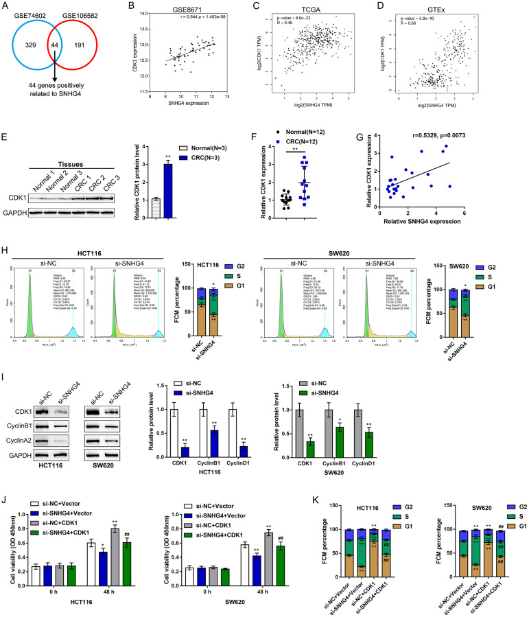
Colorectal cancer (CRC) is a prevalent malignancy worldwide. The development of genome sequencing technology has allowed the discovery that epigenetic regulation might play a critical role in CRC tumorigenesis. In the present study, we found that the long noncoding RNA (lncRNA) SNHG4 was dramatically increased in CRC tissue samples and cell lines based on both publicly available and experimental data. SNHG4 knockdown suppressed the viability and colony formation capacity of CRC cells. The expression of CDK1 was considerably increased in CRC tissue samples and cells and had a positive correlation with the expression of SNHG4 in CRC. SNHG4 silencing not only caused S phase cell cycle arrest but also significantly downregulated the CDK1, cyclin B1, and cyclin A2 protein levels in CRC cells. miR-590-3p simultaneously bound to SNHG4 and CDK1. miR-590-3p functioned to inhibit CDK1 expression. miR-590-3p overexpression exerted the same effects on the CRC cell phenotype as SNHG4 knockdown. The effects of si-SNHG4 on CRC cells were significantly reversed by anti-miR-590-3p, indicating that SNHG4 relieved the miR-590-3p-induced inhibition of CDK1 by acting as a competing endogenous RNA (ceRNA). In vivo, SNHG4 silencing inhibited subcutaneously transplanted tumor growth and decreased cell cycle marker levels, whereas miR-590-3p inhibition exerted the opposite effects. The in vivo effects of SNHG4 silencing were also reversed by miR-590-3p inhibition. The SNHG4/miR-590-3p/CDK1 axis influences the cell cycle to modulate CRC cell proliferation and subcutaneously transplanted tumor growth. Further application of this axis still requires analysis using more animal models and clinical investigations.

Keywords: CDK1; cell cycle; colorectal cancer (CRC); lncRNA SNHG4; miR-590-3p.


Conflict of interest statement

CONFLICTS OF INTEREST: The authors declare no conflicts of interest related to this study.

Figures

Figure 1
Figure 1
Expression of long noncoding RNA SNHG4 (lncRNA SNHG4) based on publicly available and experimental data. (A) The expression of SNHG4 in colorectal and normal noncancerous tissues according to the publicly available microarray expression profiles GSE8671, GSE74602, and TCGA Colon and Rectal Cancer (TCGA-CRC). (B) The expression of SNHG4 in colorectal cancer (CRC) M0-stage (nonmetastatic) and M1-stage (lymphatic or distal metastatic) tissues according to TCGA-CRC data. (C) The expression of SNHG4 in CRC microsatellite instability (MSI) and microsatellite stable (MSS) tissues according to TCGA-CRC data. (D) The expression of SNHG4 was determined in 12 CRC and normal noncancerous tissues by real-time PCR. (E) The expression of SNHG4 was determined in one normal fetal colon cell line (FHC) and five CRC cell lines (HCT8, LoVo, HCT116, SW620, and HT29) by real-time PCR. *P < 0.05, **P < 0.01, compared to FHC cells.
Figure 2
Figure 2
Effects of SNHG4 on CRC cell proliferation and metastasis. (A) SNHG4 was knocked down in HCT116 and SW620 cells by transfection of the cells with si-SNHG4 1/2/3. The transfection efficiency was validated by real-time PCR. Si-SNHG4 2 was selected for further experiments due to its better transfection efficiency. (B–C) HCT116 and SW620 cells were transfected with si-SNHG4, and (B) cell viability was examined by CCK-8 assay and (C) colony formation capacity was examined by colony formation assay. (D) HCT116 and SW620 cells were transfected with si-SNHG4, and cell invasion ability was examined by Transwell assay. *P < 0.05, **P < 0.01, compared to the si-NC group.
Figure 3
Figure 3
SNHG4 is correlated with cyclin-dependent kinase 1 (CDK1) and the CRC cell cycle. (A) Samples in GSE106582 and GSE74602 were divided into high SNHG4 expression and low SNHG4 expression groups. The correlation between SNHG4 and differentially expressed genes was analyzed to identify genes that are significantly positively correlated with SNHG4 (r > 0.40, p < 0.05); a total of 44 genes were found to be positively correlated with SNHG4 (SKA3, TOP1MT, RFC3, DSCC1, PPIL1, UBE2C, MND1, IPO4, C10orf2, CDK1, AUNIP, DDIAS, FEN1, HELLS, CDC45, PRR7, SPC25, GGCT, KIF14, RPL13, PRMT3, PROX1, CKAP2L, SLC39A10, HMMR, KIF4A, NOLC1, EXO1, MCM6, CCNF, SPDL1, NOP16, KIF20A, MCM10, EPHX4, MMP10, FJX1, NUF2, CBX2, NEK2, ANGPT2, ECT2, KIF23, and ESM1). (B–D) Correlation of the SNHG4 and CDK1 expression levels based on data from GSE8671, TCGA, and GTEx. (E) The protein levels of CDK1 in tissue samples as determined by immunoblotting. (F) The expression of CDK1 in tissue samples as determined by real-time PCR. (G) The correlation between the SNHG4 and CDK1 expression levels as determined by Pearson’s correlation analysis. (H) HCT116 and SW620 cells were transfected with si-SNHG4, and the cell cycle was examined by flow cytometry. (I) HCT116 and SW620 cells were transfected with si-SNHG4, and the protein levels of CDK1, cyclin B1, and cyclin A2 were examined. (J) HCT116 and SW620 cells were transfected with si-SNHG4 and/or CDK1 overexpression vector, and cell proliferation was examined by CCK-8 assay. (K) HCT116 and SW620 cells were transfected with si-SNHG4 and/or CDK1 overexpression vector, and cell cycle progression was examined by flow cytometry. *P < 0.05, **P < 0.01, ##P < 0.01.
Figure 4
Figure 4
miR-590-3p binds to SNHG4 and the CDK1 3'-UTR. (A) The online tools mirDIP and R programming language were used to predict miRNAs that might simultaneously target SNHG4 and the CDK1 3'-UTR, and miR-590-3p was identified. (B) The expression of miR-590-3p was determined in 12 CRC and normal noncancerous tissues by real-time PCR. (C) The expression of miR-590-3p was determined in one normal fetal colon cell line (FHC) and five CRC cell lines (HCT8, LoVo, HCT116, SW620, and HT29) by real-time PCR. (D) miR-590-3p was overexpressed or inhibited in HCT116 and SW620 cells by transfection with miR-590-3p or anti-miR-590-3p, and the effects were confirmed by real-time PCR. (E) HCT116 and SW620 cells were transfected with miR-590-3p or anti-miR-590-3p, and the mRNA levels of SNHG4 were examined by real-time PCR. (F) HCT116 and SW620 cells were transfected with miR-590-3p or anti-miR-590-3p, and the protein levels of CDK1 were examined by immunoblotting. (G–H) Luciferase reporter assays were performed by constructing luciferase reporter vectors, as described in the Materials and methods section, to validate the predicted binding of miR-590-3p to SNHG4 and the CDK1 3'-UTR. *P < 0.05, **P < 0.01, ##P < 0.01.
Figure 5
Figure 5
Effects of miR-590-3p on CRC cell proliferation and cell cycle progression. HCT116 and SW620 cells were transfected with miR-590-3p, and (A) cell viability was examined by CCK-8 assay; (B) colony formation capacity was examined by colony formation assay; (C) cell cycle progression was examined by flow cytometry; (D) the protein levels of CDK1, cyclin B1, and cyclin A2 were examined by immunoblotting. **P < 0.01, compared to the miR-NC group.
Figure 6
Figure 6
Dynamic effects of SNHG4 and its target miR-590-3p on CDK1 and CRC cell phenotype. HCT116 and SW620 cells were cotransfected with si-SNHG4 and anti-miR-590-3p, and (A) cell viability was examined by CCK-8 assay; (B) colony formation capacity was examined by colony formation assay; (C) cell cycle progression was examined by flow cytometry; and (D) the protein levels of CDK1, cyclin B1, and cyclin A2 were examined by immunoblotting. *P < 0.05, **P < 0.01, compared to the control group; #P < 0.05, ##P < 0.01, compared to the si-NC (negative control) + anti-miR-590-3p group.
Figure 7
Figure 7
Correlation of miR-590-3p expression with SNHG4 and CDK1 expression. (A–B) The correlations between miR-590-3p and SNHG4 and between miR-590-3p and CDK1 was determined using Pearson’s correlation analysis.
Figure 8
Figure 8
In vivo effects of the SNHG4/miR-590-3p axis on subcutaneously transplanted tumor growth. A subcutaneous transplantation tumor model was established in nude mice, and the mice were divided into four groups: si-NC + anti-NC group, si-SNHG4 + anti-NC group, si-NC + anti-miR-590-3p group, and si-SNHG4 + anti-miR-590-3p group. The mice received subcutaneous injections according to their group. (A–C) At day 35 of injection, the tumor size (A) was measured, the tumor volume (B) was calculated, and the tumor weight (C) was measured. (D) The protein levels of CDK1, cyclinB1, and cyclinA2 in the tumor samples were determined by immunoblotting. (E) The expression level of miR-590-3p was determined by PCR assay. (F) The expression level of SNHG4 was detected by PCR assay. *P < 0.05, **P < 0.01, compared to the si-NC + anti-NC group; ##P < 0.01, compared to si-NC + anti-miR-590-3p group.

Similar articles

Cited by

References

    1. Dekker E, Tanis PJ, Vleugels JLA, Kasi PM, Wallace MB. Colorectal cancer. Lancet. 2019; 394:1467–80. 10.1016/s0140-6736(19)32319-0 - DOI - PubMed
    1. Brenner H, Kloor M, Pox CP. Colorectal cancer. Lancet. 2014; 383:1490–502. 10.1016/s0140-6736(13)61649-9 - DOI - PubMed
    1. Nishihara R, Wu K, Lochhead P, Morikawa T, Liao X, Qian ZR, Inamura K, Kim SA, Kuchiba A, Yamauchi M, Imamura Y, Willett WC, Rosner BA, et al.. Long-term colorectal-cancer incidence and mortality after lower endoscopy. N Engl J Med. 2013; 369:1095–105. 10.1056/nejmoa1301969 - DOI - PMC - PubMed
    1. Smolle M, Uranitsch S, Gerger A, Pichler M, Haybaeck J. Current status of long non-coding RNAs in human cancer with specific focus on colorectal cancer. Int J Mol Sci. 2014; 15:13993–4013. 10.3390/ijms150813993 - DOI - PMC - PubMed
    1. Wang J, Song YX, Ma B, Wang JJ, Sun JX, Chen XW, Zhao JH, Yang YC, Wang ZN. Regulatory Roles of Non-Coding RNAs in Colorectal Cancer. Int J Mol Sci. 2015; 16:19886–919. 10.3390/ijms160819886 - DOI - PMC - PubMed

Publication types

MeSH terms