. 2021 Mar 19;13(7):9348-9372.
doi: 10.18632/aging.202719. Epub 2021 Mar 19.

miR-30c-1 encourages human corneal endothelial cells to regenerate through ameliorating senescence

Affiliations

miR-30c-1 encourages human corneal endothelial cells to regenerate through ameliorating senescence

Younghwan Bae et al. Aging (Albany NY). .

Abstract

In the present study, we studied the role of microRNA-30c-1 (miR-30c-1) on transforming growth factor beta1 (TGF-β1)-induced senescence of hCECs. hCECs were transfected by miR-30c-1 and treated with TGF-β1 to assess the inhibitory effect of miR-30c-1 on TGF-β1-induced senescence. Cell viability and proliferation rate in miR-30c-1-transfected cells was elevated compared with control. Cell cycle analysis revealed that cell abundance in S phase was elevated in miR-30c-1-treated cells compared with control. TGF-β1 increased the senescence of hCECs; however, this was ameliorated by miR-30c-1. TGF-β1 increased the size of hCECs, the ratio of senescence-associated beta-galactosidase-stained cells, secretion of senescence-associated secretory phenotype factors, the oxidative stress, and arrested the cell cycle, all of which were ameliorated by miR-30c-1 treatment. miR-30c-1 also suppressed a TGF-β1-induced depolarization of mitochondrial membrane potential and a TGF-β1 stimulated increase in levels of cleaved poly (ADP-ribose) polymerase (PARP), cleaved caspase 3, and microtubule-associated proteins 1A/1B light chain 3B II. In conclusion, miR-30c-1 promoted the proliferation of hCECs through ameliorating the TGF- β1-induced senescence of hCECs and reducing cell death of hCECs. Thus, miR-30c-1 may be a therapeutic target for hCECs regeneration.

Keywords: TGF-β; human corneal endothelial cells; miR-30c-1; proliferation; senescence.


Conflict of interest statement

CONFLICTS OF INTEREST: The authors declare no conflicts of interest.

Figures

Figure 1
Figure 1
Cell proliferation induced by miR-30c-1. (A) miR-30c level was elevated in miR-30c-1-treated cells compared with miR-control. (B) Representative images of cell shape in control and in miR-30c-1-treated cells. (C) Cell viability in control and in miR-30c-1-treated cells measured by CCK-8 assay. (D) Cell proliferation in control and in miR-30c-1-treated cells measured by BrdU proliferation assay. (E) Cell cycle analysis showing that miR-30c-1 elevated the percentage of cells with S-phase. *statistically significant.
Figure 2
Figure 2
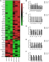
Results of RNA-sequencing data. (A) Heat map of the relative expression of differentially expressed genes. Comparison of relative expressions of proliferation-associated genes (B), interferon (IFN)-associated genes (C), transforming growth factor (TGF; (D)) caspases (E) and autophagy (F) between miR-control group and miR-30c-1 group. *statistically significant.
Figure 3
Figure 3
Oxidative stress level and mitochondrial membrane potential changed by miR-30c-1. (A) Representative images of dichlorofluorescin diacetate staining in control and miR-30c-1-treated cells. (B) DCF fluorescence intensity in control and miR-30c-1-treated cells. (C–D) Fluorescence intensity of MitoSOX probe was measured by Muse cell analyzer. (E–F) Mitochondrial membrane potential was measured using Muse® MitoPotential kit.
Figure 4
Figure 4
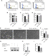
miR-30c-1 ameliorates TGF-β1-induced cell cycle arrest. (A) Cell cycle analysis was analyzed using DNA content. (B) The percentage of cells in S-phase. (C) The percentage of cells in G0/G1 phase. (D) The percentage of cells in G2/M phase. (E) Representative images of cell shape. (F) Cell size was increased by TGF-β1 treatment, which was ameliorated by miR-30c-1. (G) Representative images of yes-associated protein 1 (YAP). (H) YAP levels were quantified. (I) miR-30c-l levels were reduced in treatment with TGF-β1. *statistically significant.
Figure 5
Figure 5
miR-30c-1 ameliorates TGF-β1-induced senescence. (A) Representative images of senescence-β-galactosidase (SA-β-gal) staining. (B) The percentage of SA-β-gal positive cells was quantified. (C) Intracellular oxidative stress levels measured by MitoSOX probe. (D) Representative images of p-p38 and p38. (E) Activation of p38 was quantified. (F) Representative images of p63. (G) p63 level was quantified. *statistically significant.
Figure 6
Figure 6
Senescence-associated secretory phenotype (SASP) factors. (A–C) Interleukin-6 (IL-6; A), tumor necrosis factor-α (TNF-α; B), macrophage migration inhibitory factor (MIF; C) levels were evaluated by ELISA. (D) Representative images of immunofluorescence staining of nuclear factor kappa-light-chain-enhancer of activated B cells (NF-κB). (E) Nuclear translocation of Nf-κB was evaluated and quantified. Immunofluorescence staining of NF-κB p65 (green) and nuclear with Hoechst 33342 (blue) was performed. (F–G) SMAD2/3 levels were evaluated by western blotting. (H–I) pERK1/2 levels were evaluated by western blotting. (J) Insulin-like growth factor-1 (IGF-1) levels were evaluated by ELISA. (K) Platelet-derived growth factor-BB (PDGF-BB) were evaluated by ELISA. *statistically significant.
Figure 7
Figure 7
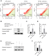
Cell death by TGF-β1 or miR-30c-1. (A) Mitochondrial membrane potential was measured by MitoPotential kit. (B–D) Cleaved caspase 9 and cleaved poly ADP ribose polymerase (PARP) levels were evaluated by western blotting and quantified. (E–F) Cleaved caspase 3 was evaluated by western blotting and quantified. *statistically significant.
Figure 8
Figure 8
Autophagy by TGF-β1 or miR-30c-1. (A) Mitochondria (red) and lysosomes (green) were staining. (B) Representative images of microtubule-associated protein 1A/1B-light chain 3 (LC3). (C) LC3I and LC3II levels were quantified. *statistically significant.

Similar articles

Cited by

References

    1. Matsubara M, Tanishima T. Wound-healing of the corneal endothelium in the monkey: a morphometric study. Jpn J Ophthalmol. 1982; 26:264–73. - PubMed
    1. Fujikawa LS, Wickham MG, Binder PS. Wound healing in cultured corneal endothelial cells. Invest Ophthalmol Vis Sci. 1980; 19:793–801. - PubMed
    1. Tamori Y, Deng WM. Compensatory cellular hypertrophy: the other strategy for tissue homeostasis. Trends Cell Biol. 2014; 24:230–7. 10.1016/j.tcb.2013.10.005 - DOI - PMC - PubMed
    1. Yang HJ, Sato T, Matsubara M, Tanishima T. [Wound healing of the corneal endothelium in the bullous keratopathy after keratoplasty]. Nippon Ganka Gakkai Zasshi. 1983; 87:701–7. - PubMed
    1. Bartel DP. MicroRNAs: genomics, biogenesis, mechanism, and function. Cell. 2004; 116:281–97. 10.1016/s0092-8674(04)00045-5 - DOI - PubMed

Publication types

MeSH terms