Review
. 2021 May 21;49(9):4831-4847.
doi: 10.1093/nar/gkab176.

Repriming DNA synthesis: an intrinsic restart pathway that maintains efficient genome replication

Affiliations
Review

Repriming DNA synthesis: an intrinsic restart pathway that maintains efficient genome replication

Lewis J Bainbridge et al. Nucleic Acids Res. .

Abstract

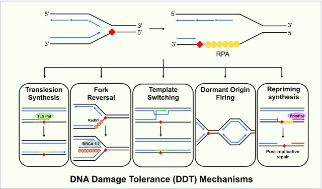
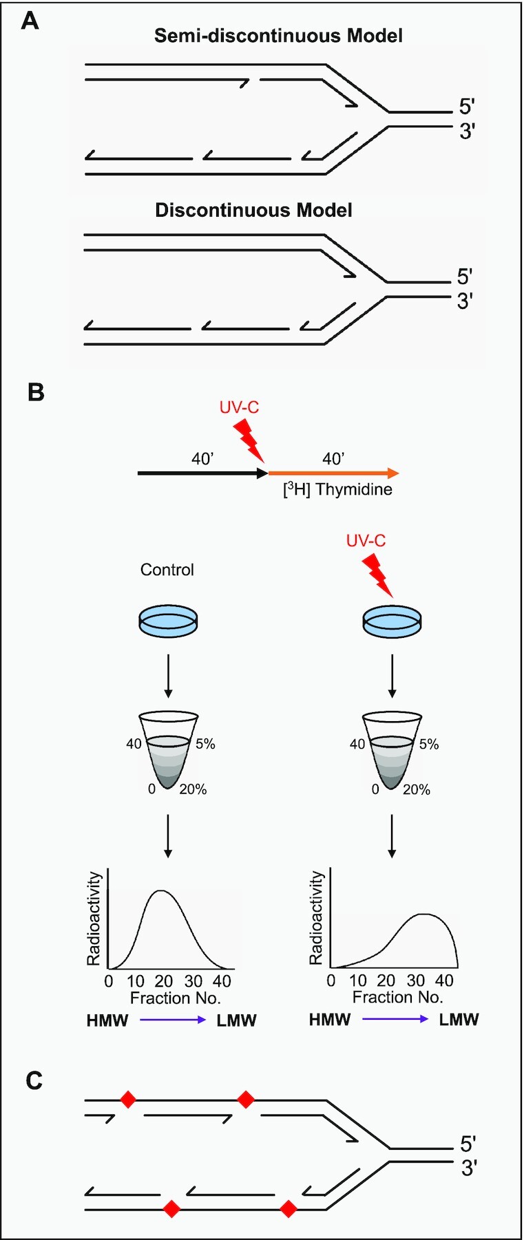
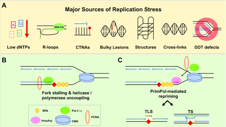
To bypass a diverse range of fork stalling impediments encountered during genome replication, cells possess a variety of DNA damage tolerance (DDT) mechanisms including translesion synthesis, template switching, and fork reversal. These pathways function to bypass obstacles and allow efficient DNA synthesis to be maintained. In addition, lagging strand obstacles can also be circumvented by downstream priming during Okazaki fragment generation, leaving gaps to be filled post-replication. Whether repriming occurs on the leading strand has been intensely debated over the past half-century. Early studies indicated that both DNA strands were synthesised discontinuously. Although later studies suggested that leading strand synthesis was continuous, leading to the preferred semi-discontinuous replication model. However, more recently it has been established that replicative primases can perform leading strand repriming in prokaryotes. An analogous fork restart mechanism has also been identified in most eukaryotes, which possess a specialist primase called PrimPol that conducts repriming downstream of stalling lesions and structures. PrimPol also plays a more general role in maintaining efficient fork progression. Here, we review and discuss the historical evidence and recent discoveries that substantiate repriming as an intrinsic replication restart pathway for maintaining efficient genome duplication across all domains of life.


Figures

Figure 1.
Figure 1.
DNA damage tolerance pathways. Obstacles on the DNA template (red diamond) block ongoing DNA replication (blue arrows) and lead to fork stalling. This leads to helicase/polymerase uncoupling, generating tracts of ssDNA, which is bound by RPA (yellow circles). DNA damage tolerance mechanisms allow DNA replication to continue in the presence of such impediments. Translesion synthesis employs specialised polymerases (green oval) to insert bases opposite damaged templating bases (orange line indicates this insertion). Fork reversal begins as the recombinase Rad51 (orange circle) replaces RPA, and, along with the recruitment of additional factors, promotes the transient remodelling of a replication fork into a stabilised ‘chicken foot’ structure to allow for lesion repair or template switching. Rad51 and BRCA 1/2 (grey oval) are factors that prevent degradation of this reversed fork structure. Template switching requires strand invasion to use the newly replicated strand as a template instead of the damaged parental strand. Dormant origin firing is activated when the replication fork slows or stalls to ensure replication is completed in a timely manner. Dormant origin firing can occur alongside the other mechanisms of DDT. Finally, repriming requires de novo primer synthesis downstream of the lesion (red arrow) from which replication can be resumed by a replicative polymerase. In vertebrate cells, this is dependent on PrimPol (pink oval), which is recruited by RPA to ssDNA.
Figure 2.
Figure 2.
Uncovering models of DNA replication. (A) In the semi-discontinuous model of DNA replication, leading strand synthesis is continuous from origin to termination and the lagging strand is synthesised in short fragments. Theoretically, if ligation is prevented, two size classes of replication intermediates would be produced: a HMW continuous leading strand and LMW fragments from the lagging strand. In the discontinuous model, both strands of DNA are synthesised as fragments and all DNA initially consists of LMW fragments. (B) The protocol used in the seminal studies of Rupp and Howard–Flanders to investigate DNA replication intermediates in bacteria following UV damage. Escherichia coli cells were grown in unlabelled medium (black arrow) before being irradiated with UV-C and transferred to media containing radioactive thymidine (orange arrow). After 40 min of labeling, the cells were collected for analysis. DNA was harvested from either control or irradiated cultures and subject to alkaline sucrose gradient centrifugation. The sedimentation showed that DNA fragments extracted from irradiated cells were significantly smaller than those from control cells. (C) The results were interpreted to indicate that gaps were present in the nascent DNA opposite the CPDs (red) induced by UV irradiation.
Figure 3.
Figure 3.
PrimPol-dependent repriming of stalled replication intermediates. (A) PrimPol-mediated repriming can assist in resolving fork stalling after many different kinds of lesions, including CTNAs, bulky lesions such as those generated by UV light, G-4 quadruplexes, R-loops, and intra/interstrand crosslinks. PrimPol can also be utilised when low dNTPs pools cause fork stalling. Additionally, the absence or loss of an alternative DDT pathway, such as fork reversal, can lead to the deployment of a PrimPol-dependent pathway. (B) Replication fork uncoupling occurs when lesions, or other sources of replication stress, transiently stall the replicative polymerase without impeding the rest of the replisome. This uncoupling generates stretches of ssDNA onto which RPA can bind. (C) PrimPol (pink oval) can be recruited to these tracts of RPA bound ssDNA to facilitate the restart of the uncoupled fork by repriming. From here, the replicative polymerase will take over to complete synthesis. The repriming depicted here occurs on the leading strand, with lagging strand machinery omitted for clarity.

Similar articles

Cited by

References

    1. Gambus A., Jones R.C., Sanchez-Diaz A., Kanemaki M., van Deursen F., Edmondson R.D., Labib K.. GINS maintains association of Cdc45 with MCM in replisome progression complexes at eukaryotic DNA replication forks. Nat. Cell Biol. 2006; 8:358–366. - PubMed
    1. Lei M. The MCM complex: its role in DNA replication and implications for cancer therapy. Curr. Cancer Drug Targets. 2005; 5:365–380. - PubMed
    1. Wohlschlegel J.A., Dwyer B.T., Dhar S.K., Cvetic C., Walter J.C., Dutta A.. Inhibition of eukaryotic DNA replication by geminin binding to Cdt1. Science. 2000; 290:2309–2312. - PubMed
    1. Evrin C., Clarke P., Zech J., Lurz R., Sun J., Uhle S., Li H., Stillman B., Speck C.. A double-hexameric MCM2-7 complex is loaded onto origin DNA during licensing of eukaryotic DNA replication. Proc. Natl. Acad. Sci. U.S.A. 2009; 106:20240–20245. - PMC - PubMed
    1. Deegan T.D., Diffley J.F.. MCM: one ring to rule them all. Curr. Opin. Struct. Biol. 2016; 37:145–151. - PubMed