

Acinar Cell Carcinoma with Morphological Change in One Month
- PMID: 33746172
- PMCID: PMC8479223
- DOI: 10.2169/internalmedicine.7121-21
Acinar Cell Carcinoma with Morphological Change in One Month
Abstract
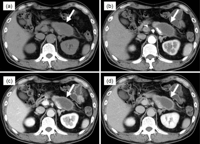
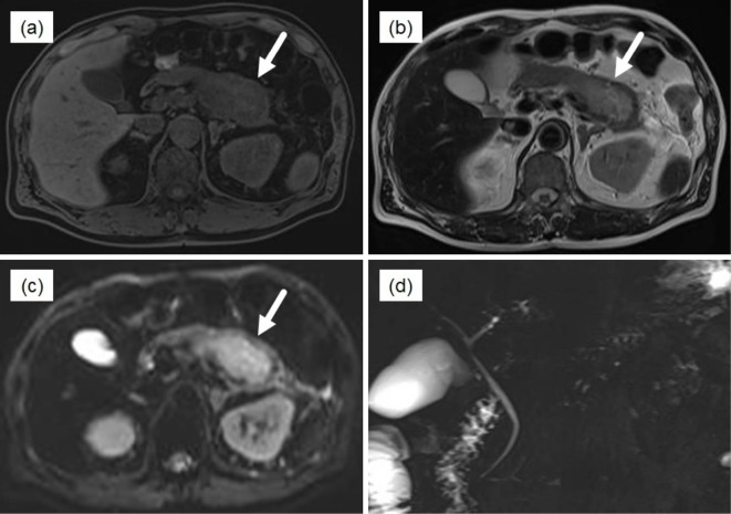
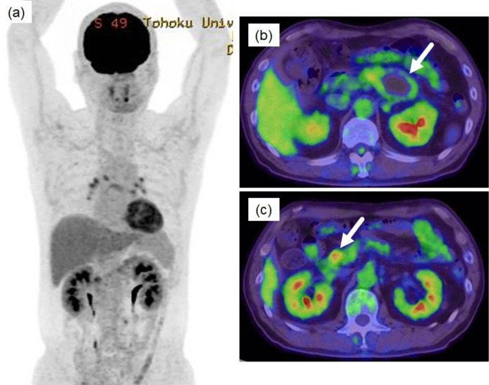
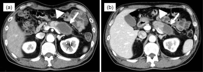
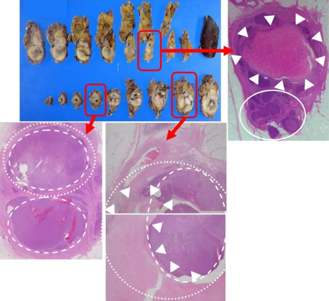
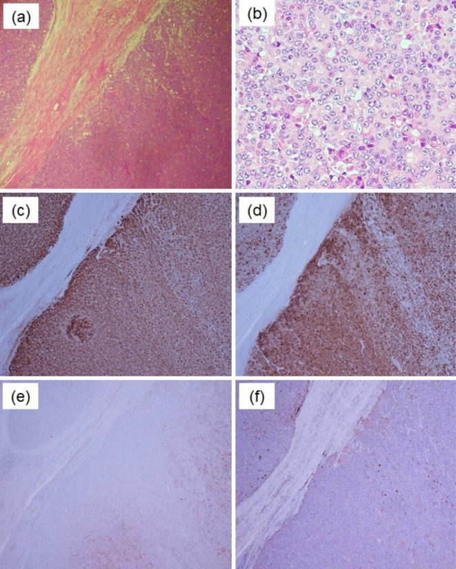
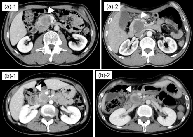
A 64-year-old man was admitted to our hospital to undergo examination of a pancreatic tumor accompanied by sudden epigastric pain. The tumor had a well-defined oval shape that was mostly less enhanced, with the exception of part of the tumor on the pancreatic head side, on contrast enhanced (CE)-CT. However, CE-CT performed one-month later revealed that the viable part of the tumor grew toward the pancreatic tail with the reduction of necrotic tissue. We performed distal pancreatectomy and the tumor was diagnosed as acinar cell carcinoma (ACC). One important characteristic of ACC is that it may develop morphological changes within a short period of time.
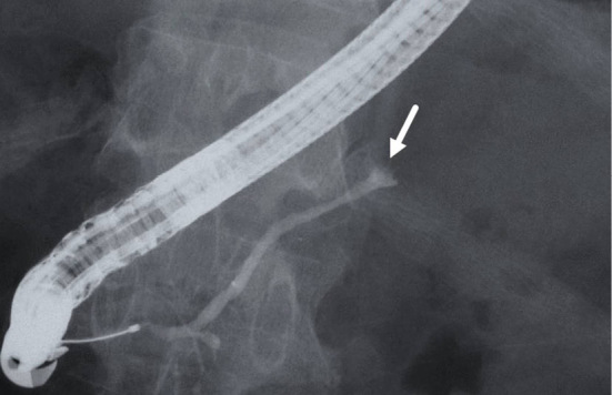
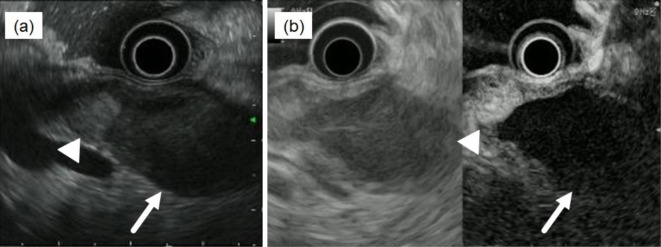
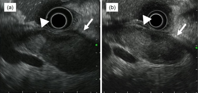
Keywords: ERCP; EUS; pancreatic cancer; pancreatic tumor.
Conflict of interest statement
Figures
Similar articles
-
[A Case Report of Acinar Cell Carcinoma of Pancreas with Extensive Intraductal Growth to the Branch - Main Pancreatic Duct].Gan To Kagaku Ryoho. 2017 Nov;44(12):1568-1570. Gan To Kagaku Ryoho. 2017. PMID: 29394704 Japanese.
-
Solid and cystic tumor of the pancreas: a case report.Acta Chir Belg. 2002 Feb;102(1):52-3. doi: 10.1080/00015458.2002.11679262. Acta Chir Belg. 2002. PMID: 11925740
-
Sudden disappearance of the blood flow in a case of pancreatic acinar cell carcinoma.Intern Med. 2014;53(22):2589-93. doi: 10.2169/internalmedicine.53.2859. Epub 2014 Nov 15. Intern Med. 2014. PMID: 25400180
-
Acinar cell carcinoma versus solid pseudopapillary tumor of the pancreas in children: a comparison of two rare and overlapping entities with review of the literature.Pediatr Dev Pathol. 2008 Sep-Oct;11(5):384-90. doi: 10.2350/07-04-0264.1. Pediatr Dev Pathol. 2008. PMID: 19006424 Review.
-
[Surgical Resection of Metachronous Hepatic Metastasis from Pancreatic Acinar Cell Carcinoma after Total Pancreatectomy].Gan To Kagaku Ryoho. 2015 Nov;42(12):2403-5. Gan To Kagaku Ryoho. 2015. PMID: 26805378 Review. Japanese.
Cited by
-
Rare Non-Neuroendocrine Pancreatic Tumours.Cancers (Basel). 2023 Apr 9;15(8):2216. doi: 10.3390/cancers15082216. Cancers (Basel). 2023. PMID: 37190144 Free PMC article. Review.
-
A case of pancreatic acinar cell carcinoma implantation in multiple branches of the pancreatic duct without main tumor continuity: a rare case report.J Gastrointest Oncol. 2024 Dec 31;15(6):2721-2727. doi: 10.21037/jgo-24-511. Epub 2024 Dec 9. J Gastrointest Oncol. 2024. PMID: 39816031 Free PMC article.
-
Comprehensive review of pancreatic acinar cell carcinoma: epidemiology, diagnosis, molecular features and treatment.Jpn J Clin Oncol. 2024 Mar 9;54(3):271-281. doi: 10.1093/jjco/hyad176. Jpn J Clin Oncol. 2024. PMID: 38109477 Free PMC article. Review.
References
-
- Kitagami H, Kondo S, Hirano S, Kawakami H, Egawa S, Tanaka M. Acinar cell carcinoma of the pancreas: clinical analysis of 115 patients from Pancreatic Cancer Registry of Japan Pancreas Society. Pancreas 35: 42-6, 2007. - PubMed
-
- Hu S, Hu S, Wang M, Wu Z, Miao F. Clinical and CT imaging features of pancreatic acinar cell carcinoma. Radiol Med 118: 723-731, 2013. - PubMed
-
- Basturk O, Zamboni G, Klimstra DS, et al. . Intraductal and papillary variants of acinar cell carcinomas: a new addition to the challenging differential diagnosis of intraductal neoplasms. Am J Surg Pathol 31: 363-370, 2007. - PubMed
-
- La Rosa S, Adsay V, Albarello L, et al. . Clinicopathologic study of 62 acinar cell carcinomas of the pancreas: insights into the morphology and immunophenotype and search for prognostic markers. Am J Surg Pathol 36: 1782-1795, 2012. - PubMed
-
- Seki M, Natori K, Kishi Y, et al. . Early image findings of acinar-endocrine carcinoma of the pancreas: a case report. Suizou 25: 693-701, 2010. (in Japanese, Abstract in English).
Publication types
MeSH terms
LinkOut - more resources
Full Text Sources
Other Literature Sources
Medical