Review
. 2021 Aug;35(8):2086-2109.
doi: 10.1038/s41433-021-01501-5. Epub 2021 Mar 21.

Ocular coloboma-a comprehensive review for the clinician

Affiliations
Review

Ocular coloboma-a comprehensive review for the clinician

Gopal Lingam et al. Eye (Lond). 2021 Aug.

Abstract

Typical ocular coloboma is caused by defective closure of the embryonal fissure. The occurrence of coloboma can be sporadic, hereditary (known or unknown gene defects) or associated with chromosomal abnormalities. Ocular colobomata are more often associated with systemic abnormalities when caused by chromosomal abnormalities. The ocular manifestations vary widely. At one extreme, the eye is hardly recognisable and non-functional-having been compressed by an orbital cyst, while at the other, one finds minimalistic involvement that hardly affects the structure and function of the eye. In the fundus, the variability involves the size of the coloboma (anteroposterior and transverse extent) and the involvement of the optic disc and fovea. The visual acuity is affected when coloboma involves disc and fovea, or is complicated by occurrence of retinal detachment, choroidal neovascular membrane, cataract, amblyopia due to uncorrected refractive errors, etc. While the basic birth anomaly cannot be corrected, most of the complications listed above are correctable to a great extent. Current day surgical management of coloboma-related retinal detachments has evolved to yield consistently good results. Cataract surgery in these eyes can pose a challenge due to a combination of microphthalmos and relatively hard lenses, resulting in increased risk of intra-operative complications. Prophylactic laser retinopexy to the border of choroidal coloboma appears to be an attractive option for reducing risk of coloboma-related retinal detachment. However, a majority of the eyes have the optic disc within the choroidal coloboma, thus making it difficult to safely administer a complete treatment.

摘要: 典型的眼组织缺损是由胚胎裂闭合不全引起的。眼组织缺损可以为偶发、遗传性 (已知或未知的基因缺陷)或与染色体异常有关。眼组织缺损多与染色体异常引起的全身异常有关。眼部表现差异多样。比如被眼眶囊肿压迫时, 其中一眼几乎无法视物、视觉功能丧失, 而另一只眼的结构和功能却丝毫不受影响。眼底的变异性则涉及到组织缺损的大小(前后和横向的范围)以及视盘和黄斑中心凹的受累程度。当组织缺损累及视盘和黄斑的中心凹时, 或合并视网膜脱离、脉络膜新生血管膜、白内障、因屈光不正引起的弱视等, 则会影响视力。虽然眼组织先天性异常无法治疗, 但上述大多数并发症在很大程度上是可以治疗的。目前, 组织缺损相关性视网膜脱离的手术治疗已经取得了良好的效果。但由于先天性小眼球和相对坚硬的晶状体, 白内障手术具有一定的挑战性, 从而增加了术中并发症的风险。脉络膜缺损边缘的预防性激光光凝治疗对于降低组织缺损相关性视网膜脱离的发病风险是一种不错的选择。然而, 大部分患眼的视盘都在脉络膜的缺损范围内, 因此很难安全的进行完整的治疗。.


Conflict of interest statement

The authors declare no competing interests.

Figures
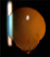
Fig. 1
Fig. 1. Slit-lamp photograph demonstrating complete iris coloboma.
Note (i) The visibility of equator of the lens inferiorly with possibly no zonules in this area (white arrow). (ii) Early vacuoles in an otherwise clear lens (yellow arrow) (color figure online).
Fig. 2
Fig. 2. Anterior segment photograph demonstrating incomplete iris coloboma.
Observe the notch in inferior pupillary border and partial thickness iris defect near iris root (arrow).
Fig. 3
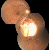
Fig. 3. Fundus photograph of choroidal coloboma.
(i) The coloboma extends above the optic disc (type 1 of Ida Mann classification). (ii) Disc coloboma merges with choroidal coloboma with disc substance hardly made out (type 6 in classification by Gopal et al.).
Fig. 4
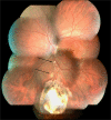
Fig. 4. Fundus photograph of choroidal coloboma.
(i) The fundus coloboma just skirts the upper disc border (type 2 of Ida Mann classification). (ii) Disc coloboma merges with fundus coloboma but upper part of disc is well delineated (type 5 of classification by Gopal et al.) (iii) Vessels meant for upper fundus emanate from lower part of the identifiable disc and smoothly go upwards across the disc. (iv) Multiple sites of emergence of inferior blood vessels from bed of the coloboma (black arrows). (v) Splash of pigment near periphery of the coloboma although there is no definable choroid or RPE (white arrows).
Fig. 5
Fig. 5. Fundus photograph of choroidal coloboma.
(i) Coloboma reaches up to lower border of disc (type 3 of Ida Mann classification). (ii) The disc is outside the coloboma and apparently normal (type 1 of the classification by Gopal et al.). However, observe the flattening of inferior border of disc. (iii) Inferior retinal blood vessels traversing within the coloboma near the border (black arrows) before supplying infero-nasal and infero temporal peripheral retina (white arrows).
Fig. 6
Fig. 6. Fundus photograph of isolated disc coloboma (type 4 Ida Mann classification).
Notice the concentric coloboma that mimics glaucomatous cupping and some degree of peripapillary atrophy.
Fig. 7
Fig. 7. Fundus photograph of choroidal coloboma.
(i) Coloboma does not reach the disc and there is normal retina above and below disc (type 5 of Ida Mann classification). (ii) Normal disc located outside the coloboma (type 1 in classification by Gopal et al). (iii) The inferior retinal blood vessel traversing the middle of the coloboma is partly obscured—hidden within the sclera (black arrows).
Fig. 8
Fig. 8. Fundus photograph of choroidal coloboma.
(i) Small coloboma restricted to inferior periphery (type 7 Ida Mann classification). (ii) Disc located outside the coloboma but dysplastic (type 2 in classification by Gopal et al). (iii) Two additional patches of non-descript chorio retinal atrophy along the line joining the disc with the coloboma-possible forme fruste lesions (black arrows).
Fig. 9
Fig. 9. Fundus photograph with choroidal coloboma.
(i) Two patches of coloboma below the optic disc and clearly separated from it. (ii) The disc is independently colobomatous with the coloboma occupying the inferior part of the disc (type 3 in classification by Gopal et al.).
Fig. 10
Fig. 10. Fundus photograph with choroidal coloboma.
Normal looking optic disc but located within the fundus coloboma (type 4 in classification by Gopal et al. and type 2 in classification by Ida Mann).
Fig. 11
Fig. 11. Collage of optical coherence tomography (OCT) images of transition zone.
A OCT demonstrating gradual transition of retina to ICM (arrow). Also notice the detachment of ICM with extension into normal retina beyond coloboma margin and the detached macula located just beyond the coloboma margin. B OCT demonstrating abrupt transition from retina to ICM (white arrow). Also observe the sudden thinning and subsequent absence of choroid at the margin of coloboma (orange arrows). C OCT demonstrating the sharp upturn of the coloboma margin (white arrows). Notice the macula close to coloboma margin. D OCT demonstrating the location of Locus minoris resistantiae (arrow). Observe the outer retinal layers turning back to merge with RPE (white arrow). E OCT demonstrating retinal detachment (orange curved arrow) extending into ICM detachment (white curved arrow). The sub-ICM space (white arrow) continues into sub-retinal space (orange arrow) due to break in Locus minoris resistantiae (thick white arrow). Inset shows location of the OCT scan. F OCT demonstrating ICM detachment with shallow retinal detachment near coloboma margin and break in Locus minoris resistantiae (orange double arrow). Observe the detached macula just beyond the coloboma margin (color figure online).
Fig. 12
Fig. 12. Fundus photograph and ICG angiogram demonstrating the type of blood vessels noted in the coloboma.
(i) Disc just outside visible limit of choroidal coloboma—normal looking but with flattened inferior border. (ii) ICG shows absence of chorio capillaris within coloboma. (iii) Black arrows delineate the retinal blood vessel that emanates from the disc, traverses through the coloboma and enters normal infero temporal retina. (iv) Absence of branches while vessel is in coloboma. (v) White arrows delineate retinal vessels emanating from bed of coloboma with some proceeding towards temporal retina. On fundus photo these can be confused as branches of the vessel marked with black arrow but ICG clearly shows them as independent vessels. Also only parts of these vessels are seen in fundus photo. (vi) Red arrows delineate retinal blood vessel that can be traced on ICG to the disc although not so obvious on fundus photo. (vii) Orange arrows delineate probably extra ocular episcleral vessels (color figure online).
Fig. 13
Fig. 13. Collage of photographs demonstrating breaks in inter-calary membrane (ICM).
A Fundus photo showing crescentic break in ICM (arrows). ICM peripheral to the break is lifted, while centrally ICM merges with the floor of coloboma. B Fundus photo showing a round hole in the anatomical macula that is involved in the coloboma (arrow). C OCT demonstrating the break in ICM (arrows) and ICM detachment. All edges of the break are lifted up.
Fig. 14
Fig. 14. Fundus photographs demonstrating peripapillary staphyloma.
A Normal disc is seen surrounded by peripapillary staphyloma. B The optic disc in this can be mistaken to be colobomatous. But close inspection clearly shows the disc to be normal (outlined by arrows). The inset shows typical optic disc coloboma for comparison.
Fig. 15
Fig. 15. Morning glory syndrome.
(i) Typical appearance of the optic disc with tuft of glial tissue at centre (white arrow), peripapillary RPE atrophy (orange arrow) and relatively straight retinal blood vessels radiating from the optic disc. (ii) Left inset shows a case of retinal detachment in an eye with morning glory syndrome. Observe the radiating folds caused by the puckering of the retina towards the disc. (iii) Right inset shows picture of ultrasonography demonstrating the funnel-shaped posterior excavation.
Fig. 16
Fig. 16. Fundus photo of macular coloboma.
Observe well delineated atrophic patch of coloboma with some pigmentation near periphery. Also observe the dense patch of medullated nerve fibres all round the optic disc.
Fig. 17
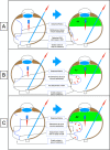
Fig. 17. Illustrations demonstrating the mechanism of RD in eyes with coloboma.
A Peripheral break present, ICM breaks absent and no dehiscence at Locus minoris resistantiae: only peripheral break contributes to the RD and RD does not extend into coloboma. B Peripheral break present, dehiscence at Locus minoris resistantiae present, ICM break absent: only peripheral break contributes to RD but RD extends into the coloboma. C ICM break present, dehiscence at Locus Minoris resistantiae present, peripheral retinal break absent: fluid spreads through ICM break to cause ICM detachment before spreading beyond coloboma to cause RD. D ICM break present, dehiscence at Locus minoris resistantiae present, peripheral retinal break present: fluid enters sub-retinal space both from peripheral retinal break and through break in ICM.
Fig. 18
Fig. 18. Illustrations demonstrating behaviour on fluid air exchange in colobomatous eyes with RD (with no attempt at sub-retinal fluid removal).
A In the presence of ICM break and dehiscence at Locus minoris resistantiae, the sub-retinal fluid is pushed into the vitreous cavity by the air and retina flattens. B In the absence of dehiscence at Locus minoris resistantiae and absence of ICM break, the sub-retinal fluid collects around coloboma and balloons the retina. There is no ICM detachment. C In the presence of dehiscence at Locus minoris resistantiae but no ICM break, fluid collects around the coloboma and balloons the retina as well as ICM.
Fig. 19
Fig. 19. Slit-lamp photograph of an eye with partial coloboma iris.
Note the dense nuclear cataract.

Similar articles

  • Ocular and systemic findings associated with optic disc colobomas.
    Berk AT, Yaman A, Saatçi AO. Berk AT, et al. J Pediatr Ophthalmol Strabismus. 2003 Sep-Oct;40(5):272-8. doi: 10.3928/0191-3913-20030901-07. J Pediatr Ophthalmol Strabismus. 2003. PMID: 14560834
  • Optic disc in fundus coloboma.
    Gopal L, Badrinath SS, Kumar KS, Doshi G, Biswas N. Gopal L, et al. Ophthalmology. 1996 Dec;103(12):2120-6; discussion 2126-7. doi: 10.1016/s0161-6420(96)30380-1. Ophthalmology. 1996. PMID: 9003347
  • Retinal detachment in a patient with microphthalmos and choroidal coloboma.
    Chen MS, Ho TC, Chang CC, Tsai TH, Hou PK. Chen MS, et al. J Formos Med Assoc. 2007 Nov;106(11):965-8. doi: 10.1016/S0929-6646(08)60069-8. J Formos Med Assoc. 2007. PMID: 18063520
  • Ocular colobomata.
    Onwochei BC, Simon JW, Bateman JB, Couture KC, Mir E. Onwochei BC, et al. Surv Ophthalmol. 2000 Nov-Dec;45(3):175-94. doi: 10.1016/s0039-6257(00)00151-x. Surv Ophthalmol. 2000. PMID: 11094243 Review.
  • [Congenital abnormalities of the optic disc].
    Denis D, Hugo J, Beylerian M, Ramtohul P, Aziz A, Matonti F, Lebranchu P. Denis D, et al. J Fr Ophtalmol. 2019 Sep;42(7):778-789. doi: 10.1016/j.jfo.2018.09.011. Epub 2019 Mar 29. J Fr Ophtalmol. 2019. PMID: 30935696 Review. French.

Cited by

  • Optic disc coloboma - A hidden masquerader.
    Tilak I, Kizhakkekara VV, Nagrajan S, Chakkaravarthy N. Tilak I, et al. Arq Bras Oftalmol. 2024 Sep 13;87(5):e20240105. doi: 10.5935/0004-2749.2024-0105. eCollection 2024. Arq Bras Oftalmol. 2024. PMID: 39292056 Free PMC article. No abstract available.
  • Congenital anomalies of lens shape.
    Bhate M, Motwani D, Murthy SI, Fernandes M. Bhate M, et al. Taiwan J Ophthalmol. 2023 Dec 20;13(4):479-488. doi: 10.4103/tjo.TJO-D-23-00076. eCollection 2023 Oct-Dec. Taiwan J Ophthalmol. 2023. PMID: 38249493 Free PMC article. Review.
  • Confounder for Optic Disc Evaluation in Glaucoma.
    Thool AR, Selukar KV, Daigavane SV. Thool AR, et al. Cureus. 2023 Feb 4;15(2):e34621. doi: 10.7759/cureus.34621. eCollection 2023 Feb. Cureus. 2023. PMID: 36891028 Free PMC article.
  • Isolated circumpapillary coloboma with normal optic disc: a rare presentation.
    Venkatesh R, Mishra P, Nahata H, Yadav NK. Venkatesh R, et al. BMJ Case Rep. 2021 Dec 27;14(12):e247353. doi: 10.1136/bcr-2021-247353. BMJ Case Rep. 2021. PMID: 34969813 Free PMC article. No abstract available.
  • Unilateral macular Coloboma: a rare finding.
    Moutei H, Bennis A, Chraibi F, Abdellaoui M, Benatiya I. Moutei H, et al. Oxf Med Case Reports. 2025 Jun 27;2025(6):omaf084. doi: 10.1093/omcr/omaf084. eCollection 2025 Jun. Oxf Med Case Reports. 2025. PMID: 40585449 Free PMC article. No abstract available.

References

    1. Duke-Elder S. Anomalous closure of embryonal cleft—typical colobomata. In: System of ophthalmology. St Louis: CV Mosby company; 1963. p. 456–88.
    1. Wilson SW, Houart C. Early steps in the development of the forebrain. Dev Cell. 2004;6:167–81. doi: 10.1016/S1534-5807(04)00027-9. - DOI - PMC - PubMed
    1. Zuber ME, Gestri G, Viczian AS, Barsacchi G, Harris WA. Specification of the vertebrate eye by a network of eye field transcription factors. Development. 2003;130:5155–67. doi: 10.1242/dev.00723. - DOI - PubMed
    1. Schwarz M, Cecconi F, Bernier G, Andrejewski N, Kammandel B, Wagner M, et al. Spatial specification of mammalian eye territories by reciprocal transcriptional repression of Pax2 and Pax6. Development. 2000;127:4325–34. doi: 10.1242/dev.127.20.4325. - DOI - PubMed
    1. Heavner W, Pevny L. Eye development and retinogenesis. Cold Spring Harb Perspect Biol. 2012;4:5–6. doi: 10.1101/cshperspect.a008391. - DOI - PMC - PubMed