

Hemorrhage and Rupture of an Unusual Benign Liver Lesion in Pregnancy: A Case Report
- PMID: 33746452
- PMCID: PMC7953012
- DOI: 10.1016/j.jceh.2020.07.004
Hemorrhage and Rupture of an Unusual Benign Liver Lesion in Pregnancy: A Case Report
Abstract
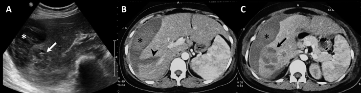
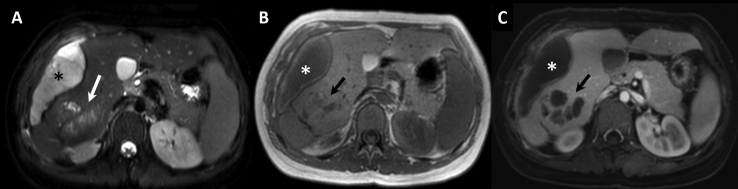
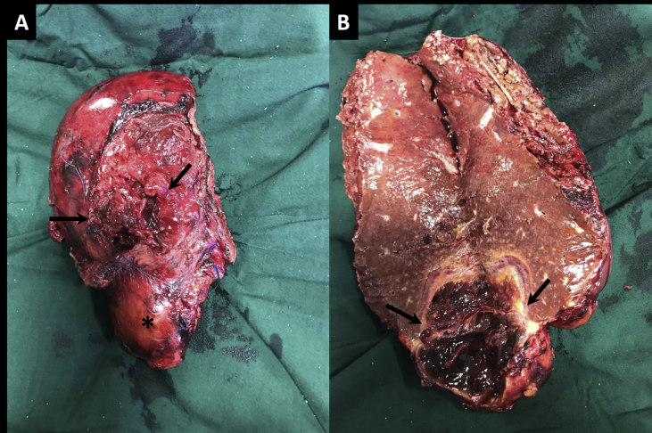
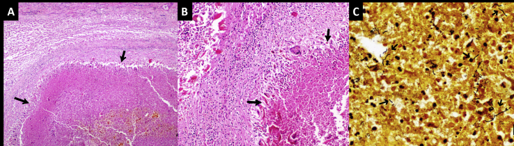
Liver rupture in pregnancy is an acute condition with significant risk to the mother and fetus. It is known to occur with tumors such as hepatic adenoma, infective causes such as abscess, granulomatous diseases, and parasitic infections, and rarely spontaneously. Most of these conditions have overlapping clinicoradiological findings, almost always requiring histopathological confirmation. We report a case of a ruptured hepatic lesion, with an unusual diagnosis of Bartonella henselae infection causing cat-scratch disease, in a 24-year-old pregnant lady.
Keywords: ALFP, Acute liver failure in pregnancy; CSD, Cat-scratch disease; CT, Computed tomography; FNH, Focal nodular hyperplasia; HCC, Hepatocellular carcinoma; HELLP, Hemolysis, elevated liver enzymes, and low platelet; IFA, Immunofluorescent assay; Ig-G, Immunoglobulin-G; MRI, Magnetic resonance imaging; PCR, Polymerase chain reaction; USG, Ultrasonography; cat-scratch disease; hemorrhagic liver lesion; liver rupture in pregnancy.
© 2020 Indian National Association for Study of the Liver. Published by Elsevier B.V. All rights reserved.
Figures
Similar articles
-
[A negleted bacteria with a case: Bartonella henselae].Mikrobiyol Bul. 2017 Jul;51(3):286-292. doi: 10.5578/mb.57321. Mikrobiyol Bul. 2017. PMID: 28929965 Turkish.
-
[A rare form of benign tumor of the liver possibly related to the use of oral contraceptives: focal pediculated nodular hyperplasia].Rev Fr Gynecol Obstet. 1985 Aug-Sep;80(8-9):621-7. Rev Fr Gynecol Obstet. 1985. PMID: 2997901 French.
-
Cat scratch disease--heterogeneous in clinical presentation: five unusual cases of an infection caused by Bartonella henselae.Klin Padiatr. 2010 Mar;222(2):73-8. doi: 10.1055/s-0029-1233488. Epub 2009 Sep 29. Klin Padiatr. 2010. PMID: 19790029
-
Hepatic adenoma and focal nodular hyperplasia.Surg Gynecol Obstet. 1991 Nov;173(5):426-31. Surg Gynecol Obstet. 1991. PMID: 1658955 Review.
-
A pediatric case of Bartonella henselae and Epstein Barr virus disease with bone and hepatosplenic involvement.Bol Med Hosp Infant Mex. 2021;78(5):467-473. doi: 10.24875/BMHIM.20000295. Bol Med Hosp Infant Mex. 2021. PMID: 34571518 Review. English.
Cited by
-
Liver imaging and pregnancy: what to expect when your patient is expecting.Insights Imaging. 2024 Feb 27;15(1):66. doi: 10.1186/s13244-024-01622-x. Insights Imaging. 2024. PMID: 38411871 Free PMC article. Review.
-
Repeated Detection of Bartonella DNA in Feline Placenta: Potential Implications for Placental and Fetal Development.Animals (Basel). 2025 Jul 11;15(14):2041. doi: 10.3390/ani15142041. Animals (Basel). 2025. PMID: 40723505 Free PMC article.
-
Bartonellas: could they cause reproductive disorders in humans?Rev Inst Med Trop Sao Paulo. 2025 Mar 3;67:e15. doi: 10.1590/S1678-9946202567015. eCollection 2025. Rev Inst Med Trop Sao Paulo. 2025. PMID: 40053001 Free PMC article. No abstract available.
References
LinkOut - more resources
Full Text Sources
Other Literature Sources
Miscellaneous
