. 2021 Feb 4;18(7):1628-1638.
doi: 10.7150/ijms.52316. eCollection 2021.

Co-culture with Endothelial Progenitor Cells promotes the Osteogenesis of Bone Mesenchymal Stem Cells via the VEGF-YAP axis in high-glucose environments

Affiliations

Co-culture with Endothelial Progenitor Cells promotes the Osteogenesis of Bone Mesenchymal Stem Cells via the VEGF-YAP axis in high-glucose environments

Peilian Wu et al. Int J Med Sci. .

Erratum in

Abstract

Patients with type 2 diabetes mellitus (T2DM) have a high risk of fracture and experience poor bone healing. In recent years, bone mesenchymal stem cells (BMSCs) and endothelial progenitor cells (EPCs) have become the most commonly used cells in cell therapy and tissue engineering. In this study, we found that high glucose levels had a negative effect on the differentiation of BMSCs and EPCs. Considering that EPCs-BMSCs sheets can provide endothelial cells and osteoblastic cells, we transplanted cell sheets into T2DM rats with bilateral skull defects. The outcomes of the in vivo study revealed that EPCs-BMSCs sheets promoted ossification, which was verified by micro-CT and immunohistochemistry (IHC) analyses. Furthermore, we detected the VEGF content in the culture supernatant using an enzyme-linked immunosorbent assay (ELISA). The results showed that the BMSCs co-cultured with EPCs presented a higher level of VEGF than other cells. To assess the differentiation and migration of BMSCs exposed to VEGF, ALP staining, scratch assay and qRT-PCR analysis were performed. In addition, we used immunofluorescence and western blotting analysis to further explore the related mechanisms. The results showed that cells cultured with VEGF had a stronger actin cytoskeleton and a greater amount of nuclear and total YAP than cells cultured without VEGF. Taken together, our results indicate that co-culture with EPCs could promote the osteogenesis of BMSCs partially via VEGF. Furthermore, YAP and F-actin play important roles in this process.

Keywords: BMSCs; EPCs; High glucose; VEGF; YAP; type 2 diabetes mellitus.


Conflict of interest statement

Competing Interests: The authors have declared that no competing interest exists.

Figures
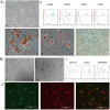
Figure 1
Figure 1
Characterization of BMSCs and EPCs. A) Isolation and differentiate of BMSCs a morphology of BNSCs,P0×100; b) Cell surface markers of BMSCs c) Alizarin red staining of BMSCs after osteogenic induction (×100; Black arrows indicate calcium deposits); d) Oil red O staining of BMSCs after adipogenic induction (×200; The black arrow indicates lipid droplets); e Alcain staining of BMSCs after chrondrogenic induction (×100). B) Isolation and identification EPCs a) morphology of epc, P1×100; b) Matrigel tubule formation experiment of EPCs (×100.Black arrow indicates tubule). c) Cell surface markers of EPCs d) Double fluorescence staining experiment of EPCs, (×200.green: FITC-UEA-1 red: DiI-Ac-LDL, yellow: fluorescence coincidence).
Figure 2
Figure 2
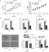
Effects of high glucose on proliferation and differentiation of BMSCs and EPCs. A) Cell viability of BMSCs and EPCs assessed by CCK-8 assay in different concentrations of glucose medium (*P<0.05); B) Effects of high glucose on differentiation of BMSCs. a) ALP ability of BMSCs (*P<0.05); b) Expressions of Runx2 mRNA (*P<0.05); c) Expressions of Osterix mRNA (*P<0.05). C) Effects of high glucose on differentiation of EPCs. a) Matrigel tubule formation experiment of EPCs in different concentrations of glucose medium (upper: ×40; lower: ×100); b) Relative protein expression of VEGF of EPCs in different concentrations of glucose medium (*P<0.05); c) Expressions of VEGF mRNA and KDR mRNA of EPCs in different concentrations of glucose medium (*P<0.05).
Figure 3
Figure 3
Establishment of cell sheets and bilateral skull defect in T2DM rats and therapeutic effect of BMSCs-EPCs sheets on skull defects. A) General observation of cell sheet and SEM observation of cell sheets (×3k). B) Establishment of T2DM rats. a) The changes in body weight between the two groups. b) The changes in glucose level between the two groups. C) Establishment of bilateral skull defect in T2DM rats a) construct 4 mm bilateral skull defects; b) implant the cell membrane into defects; c) suture. D) Micro-CT Scanning of calvarial CSD. E) Quantitative analysis of BV/TV, Tb.N, Tb.Sp, Tb.Th (*P<0.05, **P<0.001).
Figure 4
Figure 4
Immunohistochemical of bone tissue sections. A) Expression of bone marker proteins OCN in skull tissue sections from T2DM rats was detected by IHC analysis (black arrow: positive cells or intercellular substance; ×400). B) Expression of angiogenesis marker proteins VEGF in skull tissue sections from T2DM rats was detected by IHC analysis (black arrow: positive cells or intercellular substance; ×400). C) The analysis of OCN or VEGF positive cells (* P<0.05, **P<0.001).
Figure 5
Figure 5
VEGF can improve the differentiation and migration of BMSCs. A) The ALP staining of BMSCs, BMSCs+ EPCs, or BMSCs+ VEGF; B) The level of VEGF in supernatant of EPCs with or without BMSCs assessed by elisa assay (**P<0.001); C) Expressions of OCN, Osterix and Runx2 mRNA of BMSCs with or without VEGF in high concentration of glucose (*P<0.05,** P<0.001); D) Scratch experiment of BMSCs with or without VEGF in high concentration of glucose (** P<0.001); E) The ALP staining of BMSCs, BMSCs+ VEGF, or BMSCs+ VEGF+Cab; F) Expressions of VEGF, and VEGFR2 mRNA of BMSCs with or without VEGF in high concentration of glucose (*P<0.05).
Figure 6
Figure 6
VEGF improve the differentiation and migration of BMSCs may through F-actin/Yap pathway. A) Immunofluorescence images of F-actin in BMSCs with or without VEGF (scale bar = 100 um). B) Immunofluorescence images of YAP in BMSCs with or without VEGF (scale bar = 25 um). C) YAP total protein expression detected by western blotting and quantitative analysis (*P<0.05).

Similar articles

Cited by

References

    1. Liu DM, Mosialou I, Liu JM. Bone: Another potential target to treat, prevent and predict diabetes. Diabetes Obes Metab. 2018;20:1817–28. - PubMed
    1. Cancedda.R Giannoni.P, Mastrogiacomo.M. A tissue engineering approach to bone repair in large animal models and in clinical practice. Biomaterials. 2007;28:4240–50. - PubMed
    1. Murray Coleman. Impact of Diabetes Mellitus on Bone Health. International Journal of Molecular Sciences. 2019;20:4873. - PMC - PubMed
    1. Tevlin R, Seo EY, Marecic O, McArdle A, Tong X, Zimdahl B. et al. Pharmacological rescue of diabetic skeletal stem cell niches. Science Translational Medicine. 2017;9:eaag2809. - PMC - PubMed
    1. Lecka-Czernik B. Diabetes, bone and glucose-lowering agents: basic biology. Diabetologia. 2017;60:1163–9. - PMC - PubMed

MeSH terms