

Optimization of In Vivo Studies by Combining Planar Dynamic and Tomographic Imaging: Workflow Evaluation on a Superparamagnetic Nanoparticles System
- PMID: 33746630
- PMCID: PMC7953590
- DOI: 10.1155/2021/6677847
Optimization of In Vivo Studies by Combining Planar Dynamic and Tomographic Imaging: Workflow Evaluation on a Superparamagnetic Nanoparticles System
Abstract
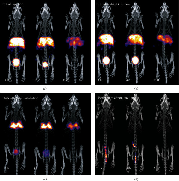
Molecular imaging holds great promise in the noninvasive monitoring of several diseases with nanoparticles (NPs) being considered an efficient imaging tool for cancer, central nervous system, and heart- or bone-related diseases and for disorders of the mononuclear phagocytic system (MPS). In the present study, we used an iron-based nanoformulation, already established as an MRI/SPECT probe, as well as to load different biomolecules, to investigate its potential for nuclear planar and tomographic imaging of several target tissues following its distribution via different administration routes. Iron-doped hydroxyapatite NPs (FeHA) were radiolabeled with the single photon γ-emitting imaging agent [99mTc]TcMDP. Administration of the radioactive NPs was performed via the following four delivery methods: (1) standard intravenous (iv) tail vein, (2) iv retro-orbital injection, (3) intratracheal (it) instillation, and (4) intrarectal installation (pr). Real-time, live, fast dynamic screening studies were performed on a dedicated bench top, mouse-sized, planar SPECT system from t = 0 to 1 hour postinjection (p.i.), and consequently, tomographic SPECT/CT imaging was performed, for up to 24 hours p.i. The administration routes that have been studied provide a wide range of possible target tissues, for various diseases. Studies can be optimized following this workflow, as it is possible to quickly assess more parameters in a small number of animals (injection route, dosage, and fasting conditions). Thus, such an imaging protocol combines the strengths of both dynamic planar and tomographic imaging, and by using iron-based NPs of high biocompatibility along with the appropriate administration route, a potential diagnostic or therapeutic effect could be attained.
Copyright © 2021 Maritina Rouchota et al.
Conflict of interest statement
The authors have no conflict of interest to declare.
Figures


Similar articles
-
On the use of superparamagnetic hydroxyapatite nanoparticles as an agent for magnetic and nuclear in vivo imaging.Acta Biomater. 2018 Jun;73:458-469. doi: 10.1016/j.actbio.2018.04.040. Epub 2018 Apr 22. Acta Biomater. 2018. PMID: 29689381
-
One pot synthesis of zwitteronic 99mTc doped ultrasmall iron oxide nanoparticles for SPECT/T1-weighted MR dual-modality tumor imaging.Colloids Surf B Biointerfaces. 2021 Jan;197:111403. doi: 10.1016/j.colsurfb.2020.111403. Epub 2020 Oct 9. Colloids Surf B Biointerfaces. 2021. PMID: 33099146
-
Inflammatory bowel disease: MR- and SPECT/CT-based macrophage imaging for monitoring and evaluating disease activity in experimental mouse model--pilot study.Radiology. 2014 May;271(2):400-7. doi: 10.1148/radiol.13122254. Epub 2014 Jan 15. Radiology. 2014. PMID: 24475849
-
The role of single-photon emission computed tomography/computed tomography in benign and malignant bone disease.Semin Nucl Med. 2006 Oct;36(4):286-94. doi: 10.1053/j.semnuclmed.2006.05.001. Semin Nucl Med. 2006. PMID: 16950146 Review.
-
Nanoparticles in practice for molecular-imaging applications: An overview.Acta Biomater. 2016 Sep 1;41:1-16. doi: 10.1016/j.actbio.2016.06.003. Epub 2016 Jun 2. Acta Biomater. 2016. PMID: 27265153 Review.
Cited by
-
Practical considerations for navigating the regulatory landscape of non-clinical studies for clinical translation of radiopharmaceuticals.EJNMMI Radiopharm Chem. 2022 Jul 19;7(1):18. doi: 10.1186/s41181-022-00168-x. EJNMMI Radiopharm Chem. 2022. PMID: 35852679 Free PMC article. Review.
References
-
- Kalishwaralal K., Luboshits G., Firer M. A. Drug Delivery Systems. New York, NY: Humana; 2019. Synthesis of gold nanoparticle: peptide–drug conjugates for targeted drug delivery; pp. 145–154. - PubMed
-
- Ahlschwede K. M., Curran G. L., Rosenberg J. T., et al. Cationic carrier peptide enhances cerebrovascular targeting of nanoparticles in Alzheimer’s disease brain. Nanomedicine: Nanotechnology, Biology and Medicine. 2019;16:258–266. - PubMed
-
- Lin M. Z., Teitell M. A., Schiller G. J. The evolution of antibodies into versatile tumor-targeting agents. Clinical Cancer Research. 2005;11(1):129–138. - PubMed
Publication types
MeSH terms
LinkOut - more resources
Full Text Sources
Other Literature Sources
Research Materials
