

Diagnostic Accuracy of 18F-FDG-PET/CT and MRI in Predicting the Tumor Response in Locally Advanced Cervical Carcinoma Treated by Chemoradiotherapy: A Meta-Analysis
- PMID: 33746650
- PMCID: PMC7943297
- DOI: 10.1155/2021/8874990
Diagnostic Accuracy of 18F-FDG-PET/CT and MRI in Predicting the Tumor Response in Locally Advanced Cervical Carcinoma Treated by Chemoradiotherapy: A Meta-Analysis
Abstract
Objective: The aim of this meta-analysis was to compare the diagnostic accuracy of 18F-FDG-PET/CT and MRI in predicting the tumor response in locally advanced cervical carcinoma (LACC) treated by chemoradiotherapy (CRT).
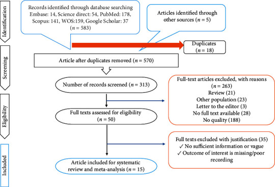
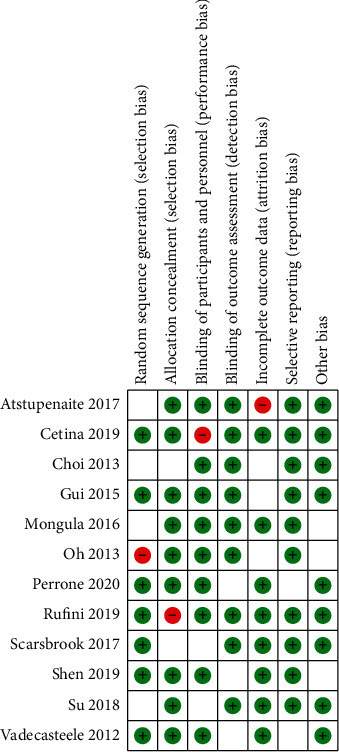
Method: This meta-analysis has been performed according to PRISMA guidelines. Systematic searches were conducted using PubMed and Embase databases for articles published from January 1, 2010, to January 1, 2020. By using the Quality Assessment of Diagnostic Accuracy Studies 2 (QUADAS-2) tool, the reviewers assessed the methodological quality scores of the selected studies. We analyzed the sensitivity, specificity, and accuracy of two diagnostic methods using Meta-DiSc 1.4 and Stata 15.
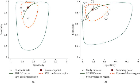
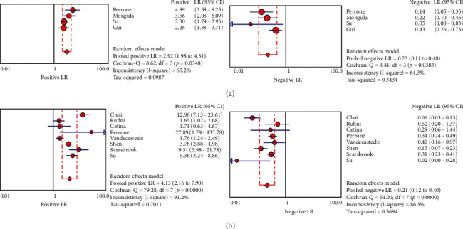
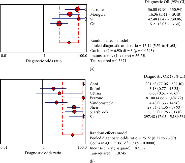
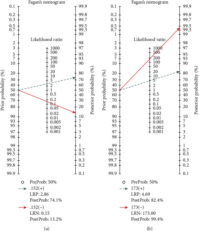
Results: An overall of 15 studies including 1132 patients were included. Sensitivities of PET/CT and MRI were 83.5% and 82.7%, while the corresponding rates for specificities were 77.8% and 68.4%, respectively. The DOR, PLR, and NLR for MRI were 15.140, 2.92, and 22.6. PET/CT had a DOR of 25.21. The PLR and NLR for PET/CT were 4.13 and 0.215, respectively. The diagnostic sensitivity and specificity of PET/CT for the detection of residual tumor were 86% and 95%, respectively. The corresponding rates for MRI were 73% and 96%, respectively. The diagnostic sensitivity and specificity of PET/CT for the detection of tumor metastases were 97% and 99%, while the corresponding rates for MRI were 31% and 98%, respectively.
Conclusion: 18F-FDG PET/CT seemed to have a better overall diagnostic accuracy in the evaluation of treatment response to chemoradiotherapy in LACC patients. MRI showed a really poor sensitivity in the detection of metastases, and PET/CT performed significantly better. However, the difference between these two methods in the detection of residual disease was not significant. More studies are needed to be conducted in order to approve that 18F-FDG PET/CT can be a standard option to assess the treatment response.
Copyright © 2021 Sharareh Sanei Sistani et al.
Conflict of interest statement
The authors declare no conflicts of interest.
Figures
Similar articles
-
Prediction of outcome using pretreatment 18F-FDG PET/CT and MRI radiomics in locally advanced cervical cancer treated with chemoradiotherapy.Eur J Nucl Med Mol Imaging. 2018 May;45(5):768-786. doi: 10.1007/s00259-017-3898-7. Epub 2017 Dec 9. Eur J Nucl Med Mol Imaging. 2018. PMID: 29222685
-
FDG PET-CT as an important diagnostic tool and prognostic marker in suspected recurrent cervical carcinoma after radiotherapy: comparison with MRI.Radiol Oncol. 2022 Nov 2;56(4):453-460. doi: 10.2478/raon-2022-0042. eCollection 2022 Dec 1. Radiol Oncol. 2022. PMID: 36317553 Free PMC article.
-
18F-FDG PET/CT and whole-body MRI diagnostic performance in M staging for non-small cell lung cancer: a systematic review and meta-analysis.Eur Radiol. 2020 Jul;30(7):3641-3649. doi: 10.1007/s00330-020-06703-1. Epub 2020 Mar 3. Eur Radiol. 2020. PMID: 32125513
-
Efficacy of qualitative response assessment interpretation criteria at 18F-FDG PET-CT for predicting outcome in locally advanced cervical carcinoma treated with chemoradiotherapy.Eur J Nucl Med Mol Imaging. 2017 Apr;44(4):581-588. doi: 10.1007/s00259-016-3537-8. Epub 2016 Oct 14. Eur J Nucl Med Mol Imaging. 2017. PMID: 27738729 Free PMC article.
-
Diagnostic Accuracy of Combined PET/CT with MRI, 18F-FDG PET/MRI, and 18F-FDG PET/CT in Patients with Oropharyngeal and Hypopharyngeal Squamous Cell Carcinoma: A Systematic Review and Meta-Analysis.Contrast Media Mol Imaging. 2021 Apr 28;2021:6653117. doi: 10.1155/2021/6653117. eCollection 2021. Contrast Media Mol Imaging. 2021. PMID: 34007251 Free PMC article.
Cited by
-
Comparing Methods to Determine Complete Response to Chemoradiation in Patients with Locally Advanced Cervical Cancer.Cancers (Basel). 2023 Dec 31;16(1):198. doi: 10.3390/cancers16010198. Cancers (Basel). 2023. PMID: 38201625 Free PMC article.
-
Post treatment imaging in patients with local advanced cervical carcinoma.Front Oncol. 2022 Oct 27;12:1003930. doi: 10.3389/fonc.2022.1003930. eCollection 2022. Front Oncol. 2022. PMID: 36465360 Free PMC article. Review.
-
Why Is Surgery Still Done after Concurrent Chemoradiotherapy in Locally Advanced Cervical Cancer in Romania?Cancers (Basel). 2024 Jan 19;16(2):425. doi: 10.3390/cancers16020425. Cancers (Basel). 2024. PMID: 38275866 Free PMC article.
-
Unraveling the Role of PET in Cervical Cancer: Review of Current Applications and Future Horizons.J Imaging. 2025 Feb 17;11(2):63. doi: 10.3390/jimaging11020063. J Imaging. 2025. PMID: 39997565 Free PMC article. Review.
-
18F-FDG PET/MRI and 18F-FDG PET/CT for the Management of Gynecological Malignancies: A Comprehensive Review of the Literature.J Imaging. 2023 Oct 13;9(10):223. doi: 10.3390/jimaging9100223. J Imaging. 2023. PMID: 37888330 Free PMC article. Review.
References
-
- Chen G., Zheng P., Gao L., Zhao J., Wang Y., Qin W. Prevalence and genotype distribution of human papillomavirus in women with cervical cancer or cervical intraepithelial neoplasia in Henan province, central China. Journal of Medical Virology. 2020;32 - PubMed
-
- Leray H., Gabiache E., Courbon F., et al. FDG-PET/CT identifies predictors of survival in patients with locally advanced cervical carcinoma and para-aortic lymph node involvement to increase treatment. Journal of Nuclear Medicine. 2020;12 - PubMed
Publication types
MeSH terms
Substances
LinkOut - more resources
Full Text Sources
Other Literature Sources
Medical
Research Materials
