

A Preliminary Study of Personalized Head CT Scan in Pediatric Patients
- PMID: 33746652
- PMCID: PMC7940743
- DOI: 10.1177/1559325820985660
A Preliminary Study of Personalized Head CT Scan in Pediatric Patients
Abstract
Objectives: In the present study, we introduced a practical approach to quantify organ-specific radiation doses and investigated whether low-dose head circumference (HC)-based protocols for non-enhanced head computed tomography (CT) could reduce organs-specific radiation dose in pediatric patients while maintaining high image quality.
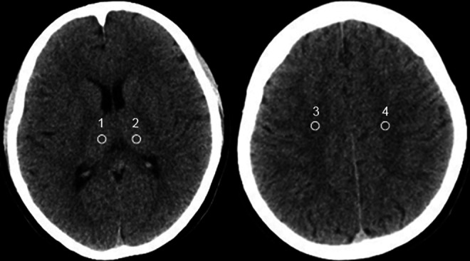
Methods: A total of 83 pediatric patients were prospectively recruited. Without limits to the HC, 15 patients were selected as a convention group (CON group) and underwent non-enhanced head CT scan with standard-dose protocols (tube current-time products of 250mAs). Low-dose group (LD group), including remaining 68 pediatrics were divided into 3 subgroups based on the HC: 54.1-57.0 cm for LD200mAs group (HC-based protocols of 200mAs), 51.1-54.0 cm for LD150mAs group (HC-based protocols of 150mAs), 48.1-51.0 cm for LD100mAs group (HC-based protocols of 100mAs). Subjective and objective image quality was evaluated and measured by 2 experienced radiologists. Radimetrics was used to calculate organs-specific radiation dose, including the brain, eye lenses, and salivary glands.
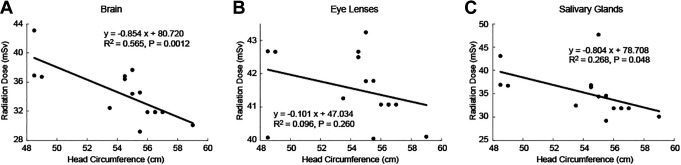
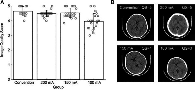
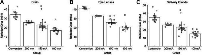
Results: In CON250mAs group, radiation doses in the brain and salivary glands were conversely correlated with HC, and pediatric patients with smaller HC received higher organs-specific radiation dose. Reducing tube current-time product from 250 to 100mAs could significantly reduce the organ-specific radiation dose. The subjective image quality score ≥ 3.0 is acceptable for diagnosis purposes. The signal to noise ratio (SNR) and the contrast to noise ratio (CNR) of bilateral thalamus and centrum semiovale in 3 LD subgroups were not statistically different compared with the CON group.
Conclusion: Our research indicated that low-dose HC-based protocols of non-enhanced head CT scan can evidently reduce the organ-specific radiation doses, while maintaining high image quality. HC can serve as a vital tool to guide personalized low-dose head CT scan for pediatric patients.
Keywords: Monte Carlo simulation; head circumference; low-dose; non-enhanced head computed tomography; pediatric; radiation dose of organs-specific; standard-dose.
© The Author(s) 2021.
Conflict of interest statement
Declaration of Conflicting Interests: The author(s) declared no potential conflicts of interest with respect to the research, authorship, and/or publication of this article.
Figures
References
-
- Sodickson A, Baeyens PF, Andriole KP, Kong CY, Barnes JA, Pandharipande PV. Recurrent CT, cumulative radiation exposure, and associated radiation-induced cancer risks from CT of adults. Radiology. 2009;251(1):175–184. - PubMed
-
- Costello JE, Cecava ND, Tucker JE, Bau JL. CT radiation dose: current controversies and dose reduction strategies. AJR Am J Roentgenol. 2013;201(6):1283–1290. - PubMed
-
- Coakley FV, Gould R, Yeh BM, Arenson RL. CT radiation dose: what can you do right now in your practice? AJR Am J Roentgenol. 2011;196(3):619–625. - PubMed
-
- Brenner DJ, Hall EJ. Computed tomography—an increasing source of radiation exposure. N Engl J Med. 2007;357(22):2277–2284. - PubMed
-
- Paolicchi F, Faggioni L, Bastiani L, et al. Optimizing the balance between radiation dose and image quality in pediatric head CT: findings before and after intensive radiologic staff training. AJR Am J Roentgenol. 2014;202(6):1309–1315. - PubMed
LinkOut - more resources
Full Text Sources
Other Literature Sources
Research Materials
