. 2021 Mar 4:14:620090.
doi: 10.3389/fnmol.2021.620090. eCollection 2021.

Complement Activation in the Central Nervous System: A Biophysical Model for Immune Dysregulation in the Disease State

Affiliations

Complement Activation in the Central Nervous System: A Biophysical Model for Immune Dysregulation in the Disease State

Nicholas Peoples et al. Front Mol Neurosci. .

Abstract

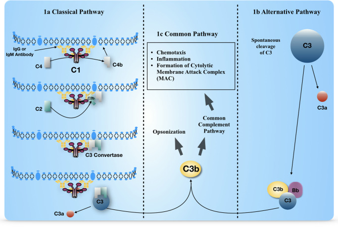
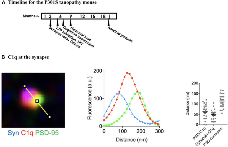
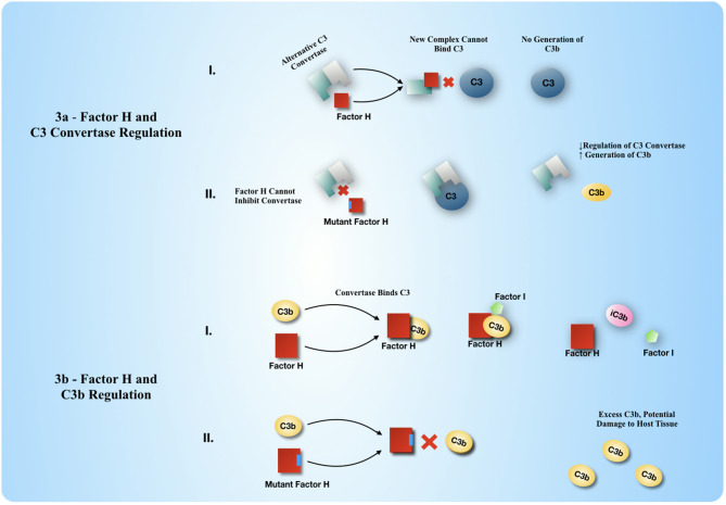
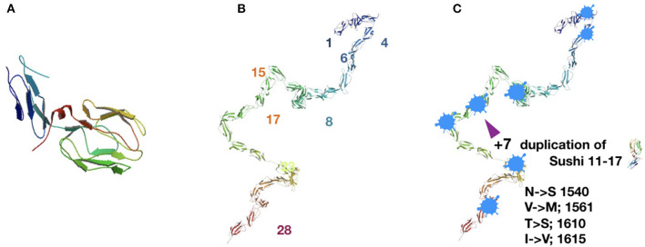
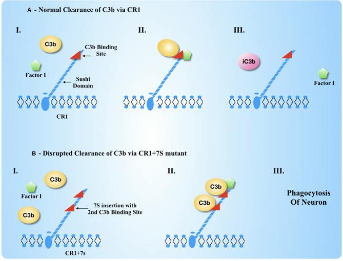
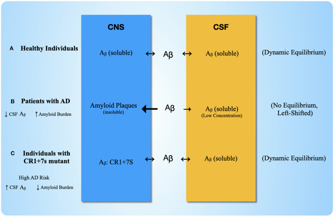
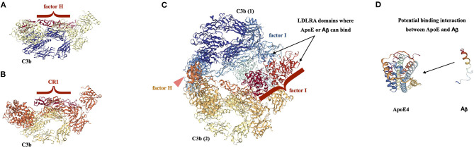
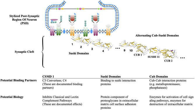
Complement, a feature of the innate immune system that targets pathogens for phagocytic clearance and promotes inflammation, is tightly regulated to prevent damage to host tissue. This regulation is paramount in the central nervous system (CNS) since complement proteins degrade neuronal synapses during development, homeostasis, and neurodegeneration. We propose that dysregulated complement, particularly C1 or C3b, may errantly target synapses for immune-mediated clearance, therefore highlighting regulatory failure as a major potential mediator of neurological disease. First, we explore the mechanics of molecular neuroimmune relationships for the regulatory proteins: Complement Receptor 1, C1-Inhibitor, Factor H, and the CUB-sushi multiple domain family. We propose that biophysical and chemical principles offer clues for understanding mechanisms of dysregulation. Second, we describe anticipated effects to CNS disease processes (particularly Alzheimer's Disease) and nest our ideas within existing basic science, clinical, and epidemiological findings. Finally, we illustrate how the concepts presented within this manuscript provoke new ways of approaching age-old neurodegenerative processes. Every component of this model is testable by straightforward experimentation and highlights the untapped potential of complement dysregulation as a driver of CNS disease. This includes a putative role for complement-based neurotherapeutic agents and companion biomarkers.

Keywords: Alzheimer's disease; C1 inhibitor; CR1; complement; factor H; neuroimmune; schizophrenia.


Conflict of interest statement

CS is an unpaid consultant to IPPIN Biomarkers and will become a shareholder of the C-Corporation should it obtain Series A financial backing. The remaining author declares that the research was conducted in the absence of any commercial or financial relationships that could be construed as a potential conflict of interest.

Figures

Figure 1
Figure 1
Elements of the classical and alternative complement pathways, separated by the unique and common aspects. (a) Early steps of the classical pathway, to show activation and generation of C3 convertase enzymatic activity. C4b (soluble) is converted to C4b (membrane bound) via a thiol ester transesterification reaction (not shown in figure). (b) Early steps of the alternative pathway to show activation and generation of C3 convertase enzymatic activity. (c) Common aspects of complement pathway activation that result in macrophage and neutrophil recruitment and formation of the membrane attack complex, a cytolytic pore on the pathogen surface.
Figure 2
Figure 2
Complement activity at the synapse in the brain of AD tauopathy mice. (A) Timeline for Alzheimer's Disease signs and symptoms to develop in P301S mouse. This mouse is analogous to a hereditary form of AD with a mutation in the tau gene that is a conversion from proline to serine at amino acid position 301. Timeline information collected from www.Alzform.org. (B) Measurements taken from micrographs of the synaptic region of the P301S mouse hippocampal CA1 region where neuronal structure is disrupted, at nine months. Data from Dejanovic et al. (2018) with permission. Fluorescence recordings from Synapsin at the presynaptic membrane, C1q, and PSD95 at the post-synaptic membrane. Analogous samples from the hippocampal CA1 region of mice without the P301S addition showed C1q staining that was barely detectable, indicating that the C1q location and increased density was due to the AD like pathology in the P301S mice.
Figure 3
Figure 3
Factor H regulates the generation of C3b and the inactivation of C3b. (a) Factor H regulates the generation of C3b by active disassembly of the C3 convertase enzyme. Factor H displaces the Bb binding partner to C3b to make a new complex with C3b that is Factor H: C3b, hence ceasing further generation of C3b. (b) Factor H regulates the level of C3b by participating in further enzymatic degradation of C3b to iC3b and other smaller protein fragments. Factor H acts as a cofactor to Factor I to bring C3b and Factor I together so that factor I may degrade C3b further through proteolysis. iC3b and smaller fragments of C3 can no longer be a part of any C3 convertase assembly, either on a cell surface or as a soluble enzyme in solution.
Figure 4
Figure 4
Complement receptor 1 biology with the basic Sushi subunit domain repeat and major and minor alleles of the gene. (A) Single Sushi domain composed of ~60 amino acids in beta sheet array, two stranded beta sheet facing three strand beta sheet, anti-parallel alignment. Major allele (left) in 83% of the population. Structure is an extracellular domain of 30 consecutive Sushi domains. Structure PDB 2Q7S, from the laboratory of SJ Perkins at UCL (Furtado et al., 2008). (B) Minor allele (right) in 11% of the population. The transmembrane domain (not shown) and basic Sushi domain subunit are unchanged. There is a seven Sushi repeat of Sushi domains 11–17, that is located directly adjacent to the original location of the duplicated Sushi domains. The binding site for C3b is located at domains 15–17, so this repeated segment of Sushi domains 11–17 includes a second binding site for C3b. (C) Inkblot; regions of predicted intrinsic disorder within the CR1 amino acid sequence. These are short regions of amino acid repeats or regions mixed with charged amino acids and hydrophobic amino acids in close proximity that create ambiguity about preference for hydrophilic environment or hydrophobic surroundings instead. Prediction programs used were: PONDR developed by Romero, Li, Dunker, Obradovic and Garner, www.pondr.com, described in Xue et al. (2010). IUPRED, www.iupred2a.elite.hu, described in Dosztányi et al. (2005).
Figure 5
Figure 5
CR1 Biology with anticipated changes due to duplicated sushi domains in CR1 biology normally and under dysregulation. (A) Under normal circumstances, CR1 acts as a cofactor to Factor I to cleave C3b into iC3b, which is released so that another C3b may be bound and cleaved. (B) Mutant CR1 with a second C3b binding site (CR1+7S) is unable to release the C3b dimer because the dimer may bind more tightly. The result is that the cell with this CR1+7S on the surface is incorrectly tagged for phagocytosis or endocytosis.
Figure 6
Figure 6
Plausible Explanation for Anomalous Clinical Data with CR1 with seven Sushi Duplication Chemical equilibrium for Aβ in three different scenarios. (A) In Scenario one, healthy individuals show a balanced chemical equilibrium, where Aβ moves into both the CNS extracellular space and the CSF at equal rates and remains soluble. (B) Scenario two shows the disruptions that occur to the equilibrium in AD patients. The Aβ in the CNS becomes insoluble in amyloid plaques, which shifts the equilibrium in favor of plaque formation in the CNS. The result is reduced levels of Aβ in the CSF. (C) Scenario three shows the equilibrium for individuals with the CR1+7s mutant. Aβ is able to bind CR1+7S, which serves as a depot for Aβ accumulation in the CNS but does not result in plaque formation, allowing continued equilibrium of Aβ into the CSF. Therefore, for those with the CR1+7S mutant, Aβ levels in the CSF will appear “normal,” but there is still an increased risk for neurodegeneration via the extra C3b binding site.
Figure 7
Figure 7
Proposed complex of CR1+7S with Additional Plasma Factors Associated with AD. Crystallographic structures from Protein Data Bank for selected RCA proteins in complex with Complement C3b. Original source for the crystallographic structures, Piet Gros laboratory (Wu et al., ; Forneris et al., ; Xue et al., 2016). These structures are used as a basis to suggest a macromolecular complex of the composition: CR1+7S: C3b: C3b: CR1+7S: ApoE. (A) Factor H: C3b complex; PDB structure, 2W11. This is a structure of the tub-like C3b structure in cream and navy, and the extended paddle-like structure of the four Sushi domains of factor H that bind to C3b resting on the lip of the tub, in brick red. (B) Complement Receptor 1: C3b complex, PDB structure 5FO9. This is a structure of the tub-like C3b structure in gold, and burnt orange, with the extended paddle—like structure of the four Sushi domains of Complement Receptor 1 that bind to C3b resting on the lip of the tub, in brick red. A comparison of the two C3b complexes where the binding partner is an RCA protein domain of consecutive Sushi domains yields many similarities. If one uses the base of the structure where there is a circular alpha helical tunnel as an orientation point, it can be seen that the C3b portion of the structures are similar to one another in pattern with similar layers of protein secondary structure. The upper surface of C3b is similar in each case and the Sushi domains of the RCA protein nestle within the middle of the structure in a nearly linear array of Sushi domains. (C) Factor H: C3b: Factor I complex; 5O32. This is a structure of two C3b domains, one in the lower half of the structure that is oriented analogously to the C3b subunits in 5a and 5b, colors yellow and gold, and a second C3b subunit in the upper half of the structure that is inverted, colors navy, slate blue. Within the middle of the C3b “sandwich” are both a Factor H Sushi string in burnt orange, and a Factor I subunit, in aqua and two shades of red. An analogous ternary structure for CR1, C3b, and factor I does not exist, but we believe that such a structure would be similar to this one, based on the extensive similarity of the Factor H: C3b and CR1: C3b structures that can be observed in (A,B). We contend that this structure is informative about the arrangement of a mega complex that might include Aβ or APP binding to the Sushi domains of CR1, quite possibly at the newly formed Sushi – Sushi interfaces made due to the seven Sushi duplication and insertion, and Apoε binding to the LDLR-A domains of factor I, shown in the two shades of red in (C). (D) ApoE and Aβ are two additional binding partners that may bind to the complex via the LDLR domains in factor I. Each of these factors may bind to an LDLR domain, or the complex of ApoE |Aβ may bind to an LDLR domain.
Figure 8
Figure 8
Post-synaptic density with CSMD. Model of CSMD protein at the protein-rich PSD. CSMD proteins are membrane bound proteins with a transmembrane domain and an extensive extracellular domain with a defined structure of 10–14 consecutive Sushi domains, then alternating CUB-Sushi domains. A stylized PSD and a CSMD protein with representative CUB and Sushi crystal structure are shown. The table below describes anticipated binding partners based on the families of proteins with CUB domains and/or Sushi domains.

Similar articles

Cited by

References

    1. Abbas A. K., Lichtman A. H., Pillai S. (2019). Basic Immunology: Functions and Disorders of the Immune System. Philidelphia, PA: Elsevier Health Sciences.
    1. Adlard P. A., Bush A. (2018). Metals and AD: how far have we come in the clinic? J. Alzheimer's Dis. 62, 1369–1379. 10.3233/JAD-170662 - DOI - PMC - PubMed
    1. Akassaglou K. (2020). The immunology of blood: connecting the dots at the neurovascular interface. Nat. Immunol. 17, 710–712. 10.1038/s41590-020-0671-z - DOI - PMC - PubMed
    1. Allen M., Kachadoorian M., Carrasquillo M. M., Karhade A., Manly L., Burgess J. D., et al. . (2015). Late-onset Alzheimer disease risk variants mark brain regulatory loci. Neurol. Genetics. 1:e15. 10.1212/NXG.0000000000000012 - DOI - PMC - PubMed
    1. Almedia J. F. F., dos Santos L. R., Trancozo M., dePaula F. (2018). Updated Meta-analysis of BIN1, CR1, MS4A6a, CLU and ABCA7 Variants in Alzheimer's disease. J. Mole Neurosci. 64, 471–477. 10.1007/s12031-018-1045-y - DOI - PubMed

LinkOut - more resources