. 2021 Mar 4:15:649262.
doi: 10.3389/fncel.2021.649262. eCollection 2021.

The Theta Rhythm of the Hippocampus: From Neuronal and Circuit Mechanisms to Behavior

Affiliations

The Theta Rhythm of the Hippocampus: From Neuronal and Circuit Mechanisms to Behavior

Angel Nuñez et al. Front Cell Neurosci. .

Abstract

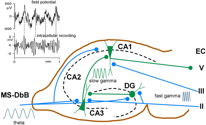
This review focuses on the neuronal and circuit mechanisms involved in the generation of the theta (θ) rhythm and of its participation in behavior. Data have accumulated indicating that θ arises from interactions between medial septum-diagonal band of Broca (MS-DbB) and intra-hippocampal circuits. The intrinsic properties of MS-DbB and hippocampal neurons have also been shown to play a key role in θ generation. A growing number of studies suggest that θ may represent a timing mechanism to temporally organize movement sequences, memory encoding, or planned trajectories for spatial navigation. To accomplish those functions, θ and gamma (γ) oscillations interact during the awake state and REM sleep, which are considered to be critical for learning and memory processes. Further, we discuss that the loss of this interaction is at the base of various neurophatological conditions.

Keywords: NMDA; cholinergic input; diagonal band of Broca; gamma oscillations; hippocampal neurons; medial septum.


Conflict of interest statement

The authors declare that the research was conducted in the absence of any commercial or financial relationships that could be construed as a potential conflict of interest.

Figures

Figure 1
Figure 1
Rhythmic medial septum-diagonal band of Broca (MS-DbB) neurons and theta (θ): in vivo recordings. (A1) Intracellular recordings of a bursting neuron with large spontaneous θ-like membrane potential oscillations (lower) in the absence of θ in the hippocampal EEG (upper). (A2) As (A1), but irregular bursting and θ. (A3) Change to higher amplitude θ and higher frequency rhythmic bursting induced by sensory stimulation (gentle stroking the animal’s back; red horizontal bar). (A4) Expanded version of part of (A3); (blue square). (B upper) Cross-correlation function (triggered by θ zero crossings, interrupted red line) of hippocampal EEG. (B lower) Cross-correlation histogram of rhythmic busting medial septum (MS) neuron activity showing strong phase locking between both activities. Note the phase advance of the neuron with θ. (C) Reset of hippocampal θ (upper) and of rhythmic bursting MS neuron (lower) triggered by reticular formation stimulation (red arrow); notice the increased amplitude of reset θ. (D) Raster plot showing successive epochs of rhythmic bursting hippocampal interneuron before, during, and after theta burst electrical stimulation of the MS. (E) Cross-correlation histogram showing entrainment by stimulation followed by phase reset (interrupted red lines). Modified from: (A) Barrenechea et al. (1995); (B,C) Gaztelu and Buño (1982); (D,E) Mamad et al. (2015).
Figure 2
Figure 2
Rhythmic CA1 pyramidal neurons and theta (θ): in vitro and in vivo recordings. (A1) Representative in vitro patch recordings showing θ–like membrane potential oscillations and action potential bursts induced by NMDA microiontophoresis at the apical dendrites of a CA1 pyramidal neuron (upper); superfision of AP5 blocked the NMDA response (lower). (A2) As (A1), but another neuron showing the effects of NMDA microiontophoresis in control ringer. (A3) Superfusion of TTX abolished action potential bursting but oscillations, NMDA- and Ca2+-spikes remained. (B1) Representative in vivo recordings of spontaneous CA1 EEG (upper) and CA1 neuron showing typical θ oscillations and single and occasional action potential bursts (lower). (B2,3) Same as (B1) in a CA3 neuron showing slow spikes riding on a sustained depolarization and firing rhythmic action potential bursts time-locked with θ oscillations; the red dots in (A3) correspond to the superimposed expanded records in panel (C). (D1) Representative in vivo tonic discharge evoked by a depolarizing current pulse applied immediately after impalement with a QX314-filled electrode. (D2) Record obtained later after impalement showing a slow putative Ca2+ spike and fewer and smaller action potentials. (D3,4) Even later, action potentials were blocked and one and two slow spikes were triggered with increasing pulse intensities (1–4 same neuron). (A) Modified from Bonansco and Buño (2003); (B,C) modified from Núñez et al. (1987); (D) modified from Nuñez and Buño (1992).
Figure 3
Figure 3
Voluntary movement and θ. (A) Phase relationship of θ with bar pressings for lateral hypothalamic (LH) electrical self-stimulation. The two superimposed averages triggered by bar pressing onsets (upper arrow) show θ waves in the pre-pressing epoch and a potential evoked by the LH self-stimulation (blue lines) followed by θ waves in the post-pressing epoch. (B1) As in (A), average triggered by bar pressing onsets (lower left arrow) but the LH self-stimulation was delayed (0.9 s) from pressings onsets (lower right arrow). Note the absence of evoked potential and pre- and post-pressing θ waves in (B1). (B2) Histogram of muscle unit firing from the left forelimb triggered by pressing onsets. (A,B) Two different rats; averages were constructed with 50 successive lever pressings. (C) Phase relationship between MS neuron discharges, step-cycles, and field θ in mice. (C1) Field θ and MS neuron action potentials (upper and lower, respectively). (C2) Forelimb movements and paw steps (upper and lower, respectively). (D) Raster plot of MS neuron firings synchronized with θ cycles and with paw steps (left and right, respectively). (A) Modified from Buño and Velluti (1977); (B) modified from Joshi and Somogyi (2020).
Figure 4
Figure 4
Place neurons and θ. (A1) Schematic diagram of rat traveling in the place field. (A2) Color coded display of place neuron firing rate in the place field. (A3,A4) Place neuron firings (red, upper) and field θ (lower). Note that the neuron displays phase precession and fires at a higher rate at a specific position in the place field. (B1) Place cell firing (green, upper) and corresponding field θ (lower) during slow locomotion (mean speed 31 cm/s). (B2) Same as (B1), but fast locomotion (mean speed 55 cm/s). The arrows indicate the time it takes the rat to cross the place field. Note that there is no change of field θ and that the neuron displays phase precession at both speeds but fires at a higher rate during the fast trial. (A) Modified from Buzsaki and Draguhn (2004); (B) modified from Geisler et al. (2007).
Figure 5
Figure 5
Schematic diagram of neuronal circuits involved in theta-gamma (θ−γ) coupling. Blue traces indicate synaptic inputs from layers II to III of the enthorynal cortex (EC) and from the MS-DbB. The dentate gyrus (DG), CA1, CA3, and EC layer V neuronal connections are also shown. θ−γ coupling in the hippocampus results from the convergence of θ inputs from the MS-DbB and fast γ from the EC. Particularly, θ−γ coupling results in CA1 by the convergence of θ inputs from the MS-DbB and slow γ from the CA3. The inset shows θ−γ interactions in the hippocampus as revealed by the CA1 field activity (upper) and band-pass filtered intracellular γ activity (lower). Note phase locking between both rhythms (θ−γ coupling) and the amplitude modulation of the intracellular γ; modified from Penttonen et al. (1998).

Similar articles

Cited by

References

    1. Abbott L. F., Nelson S. B. (2000). Synaptic plasticity: taming the beast. Nat. Neurosci. 3, 1178–1183. 10.1038/81453 - DOI - PubMed
    1. Adey W. R. (1967). Hippocwmpal states and functional relations with corticosubcortical systems in attention and learning. Prog. Brain Res. 27, 228–245. 10.1016/s0079-6123(08)63102-7 - DOI - PubMed
    1. Ahmed O. J., Mehta M. R. (2012). Running speed alters the frequency of hippocampal gamma oscillations. J. Neurosci. 32, 7373–7383. 10.1523/JNEUROSCI.5110-11.2012 - DOI - PMC - PubMed
    1. Allison S. L., Fagan A. M., Morris J. C., Head D. (2016). Spatial navigation in preclinical Alzheimer’s disease. J. Alzheimers Dis. 52, 77–90. 10.3233/JAD-150855 - DOI - PMC - PubMed
    1. Alonso A., Garcia-Austt E. (1987). Neuronal sources of theta rhythm in the entorhinal cortex of the rat. II. Phase relations between unit discharges and theta field potentials. Exp. Brain Res. 67, 502–509. 10.1007/BF00247283 - DOI - PubMed

LinkOut - more resources