. 2021 Mar 5:12:623430.
doi: 10.3389/fimmu.2021.623430. eCollection 2021.

Multi-Modal Characterization of Monocytes in Idiopathic Pulmonary Fibrosis Reveals a Primed Type I Interferon Immune Phenotype

Affiliations

Multi-Modal Characterization of Monocytes in Idiopathic Pulmonary Fibrosis Reveals a Primed Type I Interferon Immune Phenotype

Emily Fraser et al. Front Immunol. .

Abstract

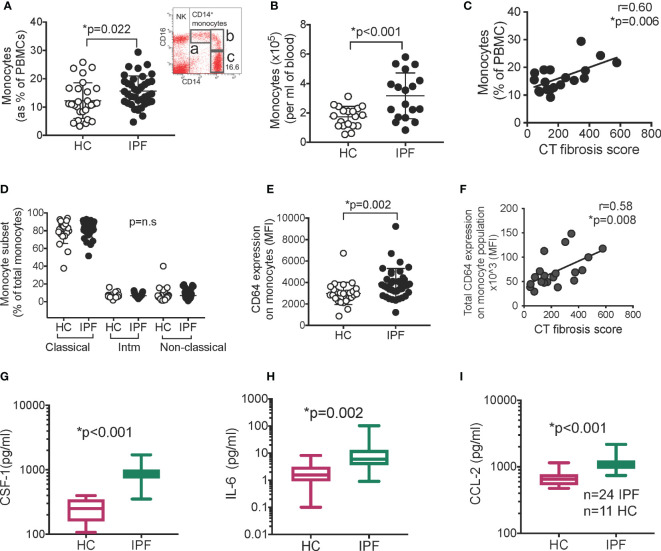
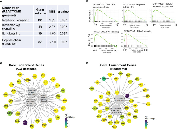
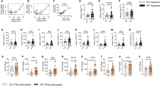
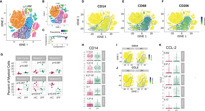
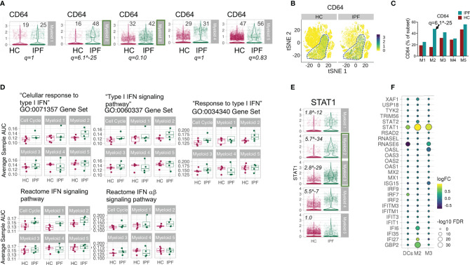
Idiopathic pulmonary fibrosis (IPF) is the most severe form of chronic lung fibrosis. Circulating monocytes have been implicated in immune pathology in IPF but their phenotype is unknown. In this work, we determined the immune phenotype of monocytes in IPF using multi-colour flow cytometry, RNA sequencing and corresponding serum factors, and mapped the main findings to amount of lung fibrosis and single cell transcriptomic landscape of myeloid cells in IPF lungs. We show that monocytes from IPF patients displayed increased expression of CD64 (FcγR1) which correlated with amount of lung fibrosis, and an amplified type I IFN response ex vivo. These were accompanied by markedly raised CSF-1 levels, IL-6, and CCL-2 in serum of IPF patients. Interrogation of single cell transcriptomic data from human IPF lungs revealed increased proportion of CD64hi monocytes and "transitional macrophages" with higher expression of CCL-2 and type I IFN genes. Our study shows that monocytes in IPF patients are phenotypically distinct from age-matched controls, with a primed type I IFN pathway that may contribute to driving chronic inflammation and fibrosis. These findings strengthen the potential role of monocytes in the pathogenesis of IPF.

Keywords: fibrosis; idiopathic pulmonary fibrosis; lung; macrophages; monocytes.


Conflict of interest statement

The authors declare that the research was conducted in the absence of any commercial or financial relationships that could be construed as a potential conflict of interest.

Figures

Figure 1
Figure 1
Monocytes and serum CSF-1, IL-6, and CCL-2 are raised in IPF. (A) Monocytes levels in IPF (n=37) vs healthy controls/HC (n=28) as % of PBMC. Inset- gating for monocytes. FACS plot was gated on all live PBMCs. “a”- non-classical monocytes, “b” –intermediate monocytes, “c” – classical monocytes; “NK” – natural killer cells (B) Monocytes as absolute number in blood (n=18). (C) Monocyte levels against amount of lung fibrosis on contemporaneous thoracic CT scans (D) Monocyte subsets in IPF and HC “Intm” – intermediate. Populations shown in (A). (E, F) Expression of CD64 (n=37) determined by flow cytometry; and correlation between CD64 expression on monocytes and CT fibrosis score (n=19). p values derived using Student t test for normally distributed data, or Mann Whitney Rank Sum test if not normally distributed. Correlation analyzed using Pearson correlation test. (G–I) Levels of CSF-1, IL-6, and CCL-2 in serum measured with Luminex technology (n=24 IPF; n=11 healthy controls, HC). Y axis in for (G–I) is expressed as log 10. Box plot is median+/1 interquartile confidence interval, whiskers show minimum and maximum values. p values calculated using Mann Whitney Rank Sum test for (G–I).
Figure 2
Figure 2
RNA sequencing of IPF monocytes reveals a Type I IFN signaling signature. (A) Analysis of differentially expressed gene list for enrichment of biologically-relevant pathway and processes using REACTOME gene sets. NES – normalized enrichment score. (B) GSEA plots showing most enriched gene sets from GO and REACTOME database. ES- Enrichment score. (C, D) Composition and expression levels of genes in the leading edge of the five gene sets from (B) expressed as log2 fold change.
Figure 3
Figure 3
Amplified response to type I IFN stimulation in monocytes from IPF. (A–C) Correlation of CD64 mRNA expression levels in IPF monocytes with ISG15 and MX1 levels; and between ISG15 and MX1 (C). (D–N) ISGs, IFNB1 and IFNAR expression in freshly isolated IPF monocytes vs HC, using qPCR. Results were expressed as relative gene expression - 1,000/2^(CT of target gene−CT of housekeeping genes). (O–V) Increased levels of ISG, STAT1, IFNA, and IFNB mRNA expression in IPF monocytes after culture in recombinant type 1 IFN for 18 h. Fold change refers to change over unstimulated values for that patient’s monocytes.
Figure 4
Figure 4
Increased number of CD64hi monocytes and CCL-2-expressing transitional macrophages in IPF lungs. (A) tSNE plot of all lung cells from lung digest of n=4 IPF explants and n=6 healthy control lungs. Broken lines outline the five subsets in the myeloid cell cluster. r-AM refers to a separate subset of cells with high cycling RNA content, some expressing CD14, CD68, and CD206 which are likely to represent embryonically-derived resident alveolar macrophages. These were referred to as “Cell Cycle” subcluster in other plots. (B) tSNE plot showing annotation of other immune cells as per Reyfman PA et al. (23). (C) Pseudotime analyses of transcriptomic states (Monocle) showing the direction of progression of states from My1 to My5. (D–F) tSNE plots showing CD14, CD68, and CD206 expression in all populations of cells from lung digest (composite of four IPF and six HC) showing progression of expression in myeloid cluster [(My1-5) in broken outline] in keeping with My1 being monocytes, My2-3, “transitional macrophages” and My4-5 being more mature macrophages. (G) Graphs showing subclusters as % of myeloid cluster. P value derived using Wilcoxon rank sum testing. (H) Violin plots for CD14 expression on each subcluster (My1-My5). Italic number refers to FDR q values comparing HC and IPF. (I, J) tSNE plots for CD14 and CCL-2, expression on all cells from lung digest, showing predominant expression in myeloid subsets (in broken outline) and increased expression in IPF compared to HC in the myeloid subsets. (K) Violin plots for CCL-2 expression on each subcluster (My1-My5). Italic number refers to FDR q values comparing HC and IPF.
Figure 5
Figure 5
Increased markers of type I IFN pathway in myeloid cells in IPF lungs. (A–C) Violin plots, tSNE and graph showing expression of CD64 in My1-5 subsets in lung digest. Numbers in plot of (A) refers to % of CD64 expressing cells in myeloid subcluster for HC and IPF patients. FDR q values applies to difference in expression between HC and IPF. (D) Average cell AUC value per sample per cluster representing enrichment of gene set in the transcriptome comparing IPF and HC samples. All p values >0.05 except for Myeloid 1 (My1) in Reactome IFN signaling pathway gene set - p=0.04 (Wilcoxon Rank Sum Test). (E) STAT1 expression in My1-5 depicted as violin plots. Italic values are FDR q values comparing HC and IPF. (F) Bubble plot reflecting expression of ISGs in My2, My3, and dendritic cells (DCs) depicted as fold change and FDR q, comparing IPF and HC, showing increased ISGs in My2 and My3 but not in DCs.

References

    1. Lederer DJ, Martinez FJ. Idiopathic Pulmonary Fibrosis. N Engl J Med (2018) 379:797–8. 10.1056/NEJMc1807508 - DOI - PubMed
    1. Martinez FJ, Collard HR, Pardo A, Raghu G, Richeldi L, Selman M, et al. . Idiopathic pulmonary fibrosis. Nat Rev Dis Primer (2017) 3:17074. 10.1038/nrdp.2017.74 - DOI - PubMed
    1. Scott MKD, Quinn K, Li Q, Carroll R, Warsinske H, Vallania F, et al. . Increased monocyte count as a cellular biomarker for poor outcomes in fibrotic diseases: a retrospective, multicentre cohort study. Lancet Respir Med (2019) 7:497–508. 10.1016/S2213-2600(18)30508-3 - DOI - PMC - PubMed
    1. Guilliams M, Mildner A, Yona S. Developmental and Functional Heterogeneity of Monocytes. Immunity (2018) 49:595–613. 10.1016/j.immuni.2018.10.005 - DOI - PubMed
    1. Misharin AV, Morales-Nebreda L, Reyfman PA, Cuda CM, Walter JM, McQuattie-Pimentel AC, et al. . Monocyte-derived alveolar macrophages drive lung fibrosis and persist in the lung over the life span. J Exp Med (2017) 214:2387. 10.1084/jem.20162152 - DOI - PMC - PubMed

Publication types

MeSH terms