Review
. 2021 Mar 4:12:649693.
doi: 10.3389/fimmu.2021.649693. eCollection 2021.

Neutrophils in the Pathogenesis of Rheumatoid Arthritis and Systemic Lupus Erythematosus: Same Foe Different M.O

Affiliations
Review

Neutrophils in the Pathogenesis of Rheumatoid Arthritis and Systemic Lupus Erythematosus: Same Foe Different M.O

Michele Fresneda Alarcon et al. Front Immunol. .

Abstract

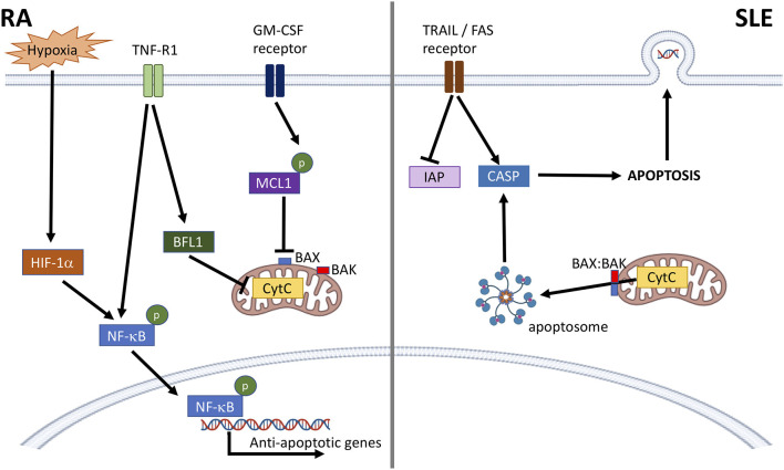
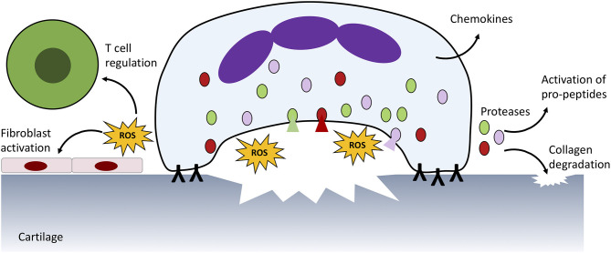
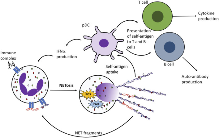
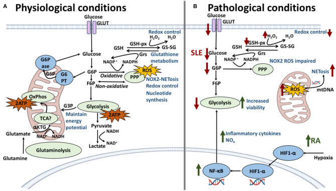
Dysregulated neutrophil activation contributes to the pathogenesis of autoimmune diseases including rheumatoid arthritis (RA) and systemic lupus erythematosus (SLE). Neutrophil-derived reactive oxygen species (ROS) and granule proteases are implicated in damage to and destruction of host tissues in both conditions (cartilage in RA, vascular tissue in SLE) and also in the pathogenic post-translational modification of DNA and proteins. Neutrophil-derived cytokines and chemokines regulate both the innate and adaptive immune responses in RA and SLE, and neutrophil extracellular traps (NETs) expose nuclear neoepitopes (citrullinated proteins in RA, double-stranded DNA and nuclear proteins in SLE) to the immune system, initiating the production of auto-antibodies (ACPA in RA, anti-dsDNA and anti-acetylated/methylated histones in SLE). Neutrophil apoptosis is dysregulated in both conditions: in RA, delayed apoptosis within synovial joints contributes to chronic inflammation, immune cell recruitment and prolonged release of proteolytic enzymes, whereas in SLE enhanced apoptosis leads to increased apoptotic burden associated with development of anti-nuclear auto-antibodies. An unbalanced energy metabolism in SLE and RA neutrophils contributes to the pathology of both diseases; increased hypoxia and glycolysis in RA drives neutrophil activation and NET production, whereas decreased redox capacity increases ROS-mediated damage in SLE. Neutrophil low-density granulocytes (LDGs), present in high numbers in the blood of both RA and SLE patients, have opposing phenotypes contributing to clinical manifestations of each disease. In this review we will describe the complex and contrasting phenotype of neutrophils and LDGs in RA and SLE and discuss their discrete roles in the pathogenesis of each condition. We will also review our current understanding of transcriptomic and metabolomic regulation of neutrophil phenotype in RA and SLE and discuss opportunities for therapeutic targeting of neutrophil activation in inflammatory auto-immune disease.

Keywords: NETs; immunometabolism; low density granulocytes; neutrophils; rheumatoid arthritis; systemic lupus erythematosus.


Conflict of interest statement

The authors declare that the research was conducted in the absence of any commercial or financial relationships that could be construed as a potential conflict of interest.

Figures

Figure 1
Figure 1
Dysregulation of neutrophil apoptosis in RA and SLE. In RA, anti-apoptotic factors, such as cytokines and hypoxia activate NF-κB and prevent mitochondrial cytochrome c (CytC) driven apoptosis through activation of BFL1, and stabilization of MCL1. This prevents BAX:BAK pore formation, CytC leakage and formation of the apoptosome. In SLE, activation of death receptors (e.g., TRAIL, FAS receptors) lowers levels of cellular inhibitors of apoptosis (IAP) and activates caspase-8 (CASP). BAX:BAK pore formation in the mitochondrial membrane releases CytC leading to apoptosome formation and activation of CASP-9 and CASP-3 leading to apoptosis. Nuclear antigens, including DNA, are also expressed at the plasma membrane.
Figure 2
Figure 2
Neutrophil ROS production and protease release damages host tissue. In RA, immune complexes on the surface of the joint activate ROS production and protease release from granules (shaded red, green, and pink) which damages underlying cartilage and activates neighbouring immune cells and fibroblasts. Proteases also activate pro-peptides, such as cytokines produced by neutrophils and other infiltrating immune cells. A similar process is responsible for damage to blood vessels in SLE.
Figure 3
Figure 3
NET production in RA and SLE drives the auto-immune response. Fragments of DNA and proteins (including histones, MPO, elastase, HMGB1, LL37) are taken up by plasmacytoid dendritic cells (pDCs) and presented to auto-reactive B- and T-cells, leading to production of cytokines including interferon alpha (IFNα) and autoantibodies. NET fragments, including oxidized DNA (shown in orange) may also activate NET production by neighboring neutrophils.
Figure 4
Figure 4
Neutrophil metabolic pathways and the effect of their dysregulation in SLE and RA. (A) In physiological conditions, glycolysis is the main energy producing pathway utilized by neutrophils; intermediate metabolites G6P and F6P are starter molecules for the pentose phosphate pathway (PPP), important in redox control, NOX2-dependent NETosis and ROS production. The oxidative stage of PPP produces NADPH which is used in the glutathione metabolism to reduce glutathione providing further redox capacity. Other energy producing pathways have been described, previously thought to be non-functional, such as the TCA cycle which connects glutaminolysis pathway and further regulates NOX2 complex by generating the reducing equivalents of NADPH. (B) In SLE and RA dysregulation of these pathways is responsible for the conditions observed at the cellular level. In SLE (red arrows), a lower expression of glucose transporters is met with lower levels of intracellular glucose, diminishing the energy output by glycolysis and compromising cellular viability. Furthermore, a decrease of G6P intermediates correlates with lower levels of NOX2-dependent ROS and decreased redox capacity. NOX2-independent ROS production in mitochondria is amplified in SLE neutrophils with increasing release of NETs containing mitochondrial DNA. In RA (green arrows), the inflammatory environment and hypoxic conditions increase the expression of HIF1-α which upregulates key glycolytic enzymes increasing energy production and viability. HIF1-α also is an upstream regulator of NF-κB which increases pro-inflammatory cytokine production therefore maintaining the inflammatory environment.

Similar articles

Cited by

References

    1. Bengtsson AA, Ronnblom L. Systemic lupus erythematosus: still a challenge for physicians. J Intern Med. (2017) 281:52–64. 10.1111/joim.12529 - DOI - PubMed
    1. Isenberg DA, Manson JJ, Ehrenstein MR, Rahman A. Fifty years of anti-ds DNA antibodies: are we approaching journey's end? Rheumatology (Oxford). (2007) 46:1052–6. 10.1093/rheumatology/kem112 - DOI - PubMed
    1. Ward MM. Premature morbidity from cardiovascular and cerebrovascular diseases in women with systemic lupus erythematosus. Arthritis Rheum. (1999) 42:338–46. 10.1002/1529-0131(199902)42:2<338::AID-ANR17>3.0.CO;2-U - DOI - PubMed
    1. Lisnevskaia L, Murphy G, Isenberg D. Systemic lupus erythematosus. Lancet. (2014) 384:1878–88. 10.1016/S0140-6736(14)60128-8 - DOI - PubMed
    1. Davidson A. What is damaging the kidney in lupus nephritis? Nat Rev Rheumatol. (2016) 12:143–53. 10.1038/nrrheum.2015.159 - DOI - PMC - PubMed

Publication types

MeSH terms