. 2021 Mar 3:12:629045.
doi: 10.3389/fpls.2021.629045. eCollection 2021.

Retracing Storage Polysaccharide Evolution in Stramenopila

Affiliations

Retracing Storage Polysaccharide Evolution in Stramenopila

Malika Chabi et al. Front Plant Sci. .

Abstract

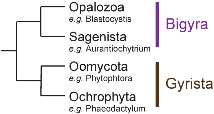
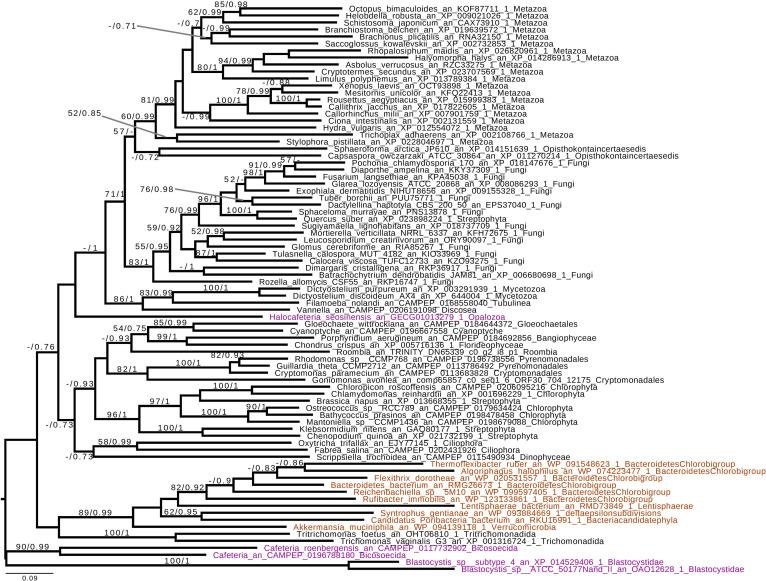
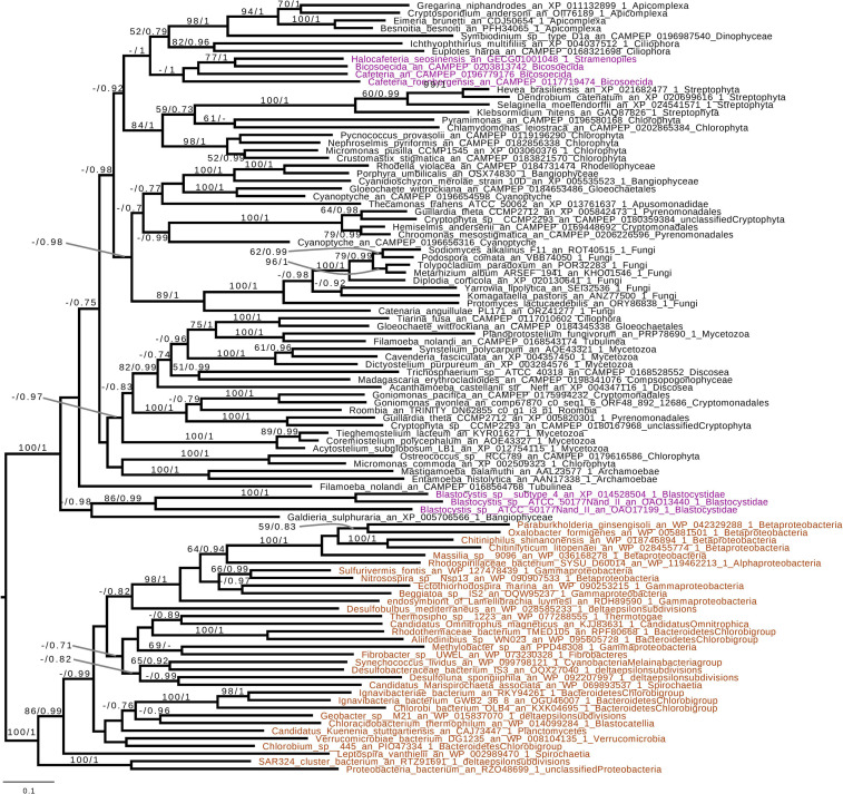
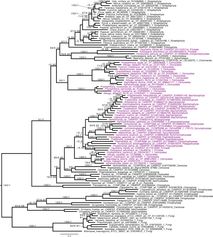
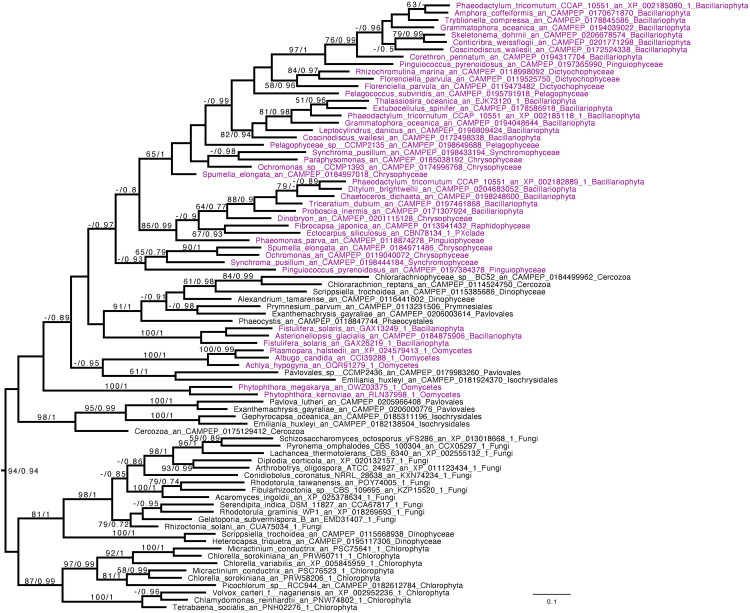
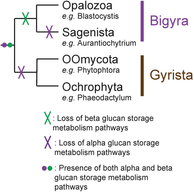
Eukaryotes most often synthesize storage polysaccharides in the cytosol or vacuoles in the form of either alpha (glycogen/starch)- or beta-glucosidic (chrysolaminarins and paramylon) linked glucan polymers. In both cases, the glucose can be packed either in water-soluble (glycogen and chrysolaminarins) or solid crystalline (starch and paramylon) forms with different impacts, respectively, on the osmotic pressure, the glucose accessibility, and the amounts stored. Glycogen or starch accumulation appears universal in all free-living unikonts (metazoa, fungi, amoebozoa, etc.), as well as Archaeplastida and alveolata, while other lineages offer a more complex picture featuring both alpha- and beta-glucan accumulators. We now infer the distribution of these polymers in stramenopiles through the bioinformatic detection of their suspected metabolic pathways. Detailed phylogenetic analysis of key enzymes of these pathways correlated to the phylogeny of Stramenopila enables us to retrace the evolution of storage polysaccharide metabolism in this diverse group of organisms. The possible ancestral nature of glycogen metabolism in eukaryotes and the underlying source of its replacement by beta-glucans are discussed.

Keywords: CAZy; glycogen; laminarin; metabolism; polysaccharide; stramenopila.


Conflict of interest statement

The authors declare that the research was conducted in the absence of any commercial or financial relationships that could be construed as a potential conflict of interest.

Figures

FIGURE 1
FIGURE 1
Schematic representation of the Stramenopila evolution tree.
FIGURE 2
FIGURE 2
Phylogenetic analysis of branching enzyme, Glycoside Hydrolase family 13 subfamily 8 (GH13_8). The tree displayed is midpoint rooted and represents the consensus tree obtained with Phylobayes 4.1 with ML bootstrap values drawn from 100 bootstraps repetition with IQTREE (left) and Bayesian posterior probabilities (right) mapped onto the nodes. Bootstrap values >50% are shown, while only posterior probabilities >0.6 are shown. The scale bar shows the inferred number of amino acid substitutions per site. Sequences are highlighted in purple for Stramenopila, brown for Bacteria, while everything else is in black. Sequence names are composed of the organism name, the accession number, and their clades. We can see that Blastocystis and Cafeteria group together, however, with a low bootstrap and posterior probabilities. In addition, Halocafeteria was not found inside a monophyletic group, but was found among other eukaryotes.
FIGURE 3
FIGURE 3
Phylogenetic analysis of glycogen phosphorylase (GT35 family). The tree displayed is midpoint rooted and represents the consensus tree obtained with Phylobayes 4.1 with ML bootstrap values drawn from 100 bootstraps repetition with IQTREE (left) and Bayesian posterior probabilities (right) mapped onto the nodes. Bootstrap values >50% are shown, while only posterior probabilities >0.6 are shown. The scale bar shows the inferred number of amino acid substitutions per site. Sequences are highlighted in purple for Stramenopila, brown for Bacteria, while everything else is in black. Sequence names are composed of the organism name, the accession number, and their clades. We can see that Bicosoecida, Halocafeteria, and Cafeteria group together, with a high posterior probability (pp = 1). However, Blastocystis was not found inside a monophyletic group with Stramenopila but was found among other eukaryotes. The Blastocystis position is probably due to the fast-evolving sequence in this organism as it has been observed in several studies (Eme et al., 2017; Moreira and López-García, 2017).
FIGURE 4
FIGURE 4
Phylogenetic analysis of GT48. The tree displayed is midpoint rooted and represents the consensus tree obtained with Phylobayes 4.1 with ML bootstrap values drawn from 100 bootstraps repetition with IQTREE (left) and Bayesian posterior probabilities (right) mapped onto the nodes. Bootstrap values >50% are shown, while only posterior probabilities >0.6 are shown. The scale bar shows the inferred number of amino acid substitutions per site. Sequences are highlighted in purple for Stramenopila, while everything else is in black. Sequence names are composed of the organism name, the accession number, and their clades. We can observe a strongly supported group with all Gyrista (BS = 99, pp = 1), with inside the expected topology based on Stramenopila phylogeny. In addition, among those sequences we find the characterized enzyme from P. tricornutum. Then, this group of sequence is likely to be the one involved in laminarin biosynthesis. Moreover, we can find close to this Gyrista group several sequences from both Haptophyta and Cercozoa (BS = 79, pp = 0.96), probably also involved in storage polysaccharide metabolism.
FIGURE 5
FIGURE 5
Phylogenetic analysis of GH16_2. The tree displayed is manually rooted according to the topology obtained in Supplementary Figure S3. It represents the consensus tree obtained with Phylobayes 4.1 with ML bootstrap values drawn from 100 bootstraps repetition with IQTREE (left) and Bayesian posterior probabilities (right) mapped onto the nodes. Bootstrap values (BS) >50 are shown, while only posterior probabilities (pp) >0.6 are shown. The scale bar shows the inferred number of amino acid substitutions per site. Sequences are highlighted in purple for Stramenopila, while everything else is in black. Sequences names are composed of the organism name, the accession number, and their clades. GH16_2 from the Stramenopila group together with sequences from Haptista and Cercozoa with a BS = 94 and a pp = 0.94, mirroring the topology from the GT48 phylogenetic tree.
FIGURE 6
FIGURE 6
Schematic representation of the Stramenopila evolution tree with the hypothesis defended here with the presence of both alpha- and beta-glucan storage polysaccharide metabolisms in the ancestor, represented by the purple and green dot, respectively. Their respective loss is indicated by a cross on the branch: in Bigyra, the loss of beta-glucan metabolism is indicated by a green cross, while alpha-glucan metabolism has been lost twice in Gyrista lineages as well as in Sagenista (purple cross).

References

    1. Almagro Armenteros J. J., Salvatore M., Emanuelsson O., Winther O., von Heijne G., Elofsson A., et al. (2019). Detecting sequence signals in targeting peptides using deep learning. Life Sci. Alliance 2 e201900429. 10.26508/lsa.201900429 - DOI - PMC - PubMed
    1. Ball S., Colleoni C., Cenci U., Raj J. N., Tirtiaux C. (2011). The evolution of glycogen and starch metabolism in eukaryotes gives molecular clues to understand the establishment of plastid endosymbiosis. J. Exp. Bot. 62 1775–1801. 10.1093/jxb/erq411 - DOI - PubMed
    1. Baurain D., Brinkmann H., Petersen J., Rodriguez-Ezpeleta N., Stechmann A., Demoulin V., et al. (2010). Phylogenomic evidence for separate acquisition of plastids in cryptophytes, haptophytes, and stramenopiles. Mol. Biol. Evol. 27 1698–1709. 10.1093/molbev/msq059 - DOI - PubMed
    1. Bowen S., Wheals A. (2004). Incorporation of Sed1p into the cell wall of involves. FEMS Yeast Res. 4 731–735. 10.1016/j.femsyr.2004.02.006 - DOI - PubMed
    1. Burki F., Kaplan M., Tikhonenkov D. V., Zlatogursky V., Minh B. Q., Radaykina L. V., et al. (2016). Untangling the early diversification of eukaryotes: a phylogenomic study of the evolutionary origins of Centrohelida, Haptophyta and Cryptista. Proc. R. Soc. B Biol. Sci. 283 20152802. 10.1098/rspb.2015.2802 - DOI - PMC - PubMed

LinkOut - more resources