. 2021 Mar 2;12(5):450-474.
doi: 10.18632/oncotarget.27895.

Development of a ghrelin receptor inverse agonist for positron emission tomography

Affiliations

Development of a ghrelin receptor inverse agonist for positron emission tomography

Ralf Bergmann et al. Oncotarget. .

Abstract

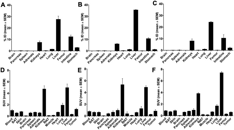
Imaging of Ghrelin receptors in vivo provides unique potential to gain deeper understanding on Ghrelin and its receptors in health and disease, in particular, in cancer. Ghrelin, an octanoylated 28-mer peptide hormone activates the constitutively active growth hormone secretagogue receptor type 1a (GHS-R1a) with nanomolar activity. We developed novel compounds, derived from the potent inverse agonist K-(D-1-Nal)-FwLL-NH2 but structurally varied by lysine conjugation with 1,4,7-triazacyclononane,1-glutaric acid-4,7-acetic acid (NODAGA), palmitic acid and/or diethylene glycol (PEG2) to allow radiolabeling and improve pharmacokinetics, respectively. All compounds were tested for receptor binding, potency and efficacy in vitro, for biodistribution and -kinetics in rats and in preclinical prostate cancer models on mice. Radiolabeling with Cu-64 and Ga-68 was successfully achieved. The Cu-64- or Ga-68-NODAGA-NH-K-K-(D-1-NaI)-F-w-L-L-NH2 radiotracer were specifically accumulated by the GHS-R1a in xenotransplanted human prostate tumor models (PC-3, DU-145) in mice. The tumors were clearly delineated by PET. The radiotracer uptake was also partially blocked by K-(D-1-Nal)-FwLL-NH2 in stomach and thyroid. The presence of the GHS-R1a was also confirmed by immunohistology. In the arterial rat blood plasma, only the original compounds were found. The Cu-64 or Ga-68-NODAGA-NH-K-K-(D-1-NaI)-F-w-L-L-NH2 radiolabeled inverse agonists turned out to be potent and safe. Due to their easy synthesis, high affinity, medium potency, metabolic stability, and the suitable pharmacokinetic profiles, they are excellent tools for imaging and quantitation of GHS-R1a expression in normal and cancer tissues by PET. These compounds can be used as novel biomarkers of the Ghrelin system in precision medicine.

Keywords: cancer; copper-64; growth hormone secretagogue receptor (GHS-R); prostate cancer; small animal imaging.


Conflict of interest statement

CONFLICTS OF INTEREST Authors have no conflicts of interest to declare.

Figures

Figure 1
Figure 1. Synthesis of peptides 3–18.
All peptides were synthesized on solid support using the Fmoc/tBu strategy and Mtt protecting groups for orthogonal side-chain modifications.
Figure 2
Figure 2. Radio-HPLC of rat blood plasma.
Radio-HPLC of rat blood plasma at 0 and 60 or 120 minutes after single intravenous injection of the 68Ga-radiotracers (AC) 9b, 10b and 15b and the 64Cu-radiotracers (DF) 9c, 10c and 15c.
Figure 3
Figure 3. Biodistribution of 68Ga-radiotracers in healthy rats.
Biodistribution of 68Ga-radiotracers 9b, 10b and 15b in healthy male Wistar rats after single intravenous injection at 5 and 60 min, the activity amounts in selected organs are expressed as percent of the injected dose (A, % ID) and the activity concentrations in the tissues are given as standard uptake values (B, SUV).
Figure 4
Figure 4. Biodistribution of 64Cu-radiotracers in healthy rats.
Biodistribution of 64Cu-radiotracers 9c, 10c and 15c in healthy male Wistar rats after single intravenous injection at 5 and 60 min, the activity amounts in selected organs are expressed as percent of the injected dose (A, % ID) and the activity concentrations in the tissues are given as standard uptake values (B, SUV).
Figure 5
Figure 5. PET studies of 68Ga- and 64Cu-labeled radiotracers in healthy rats.
PET studies of radiotracers 9b, 9c, 10b, 10c, 15b and 15c in Wistar rats. The images represent the maximum intensity projections of the radiotracer distribution 1 h after injection. The figures show the time-activity curves in the clearly visible and definable organs (dynamic measurement over one hour). Values are given as SUVmean.
Figure 6
Figure 6. Biodistribution of 10c in DU-145 tumor bearing NMRI nu/nu mice 60 min p.i.
Biodistribution of 10c in DU-145 tumor bearing NMRI nu/nu mice 60 min p.i. (A, D) Control. (B, E) Blocked with 1 mg/kg body weight Ghrelin. (C, F) Blocked with 1 mg/kg body weight KKD. Values are presented as % ID and SUV as mean ± SEM of three animals.
Figure 7
Figure 7. Biodistribution of 10c in PC-3 tumor bearing NMRI nu/nu mice 60 min p.i.
Biodistribution of 10c in PC-3 tumor bearing NMRI nu/nu mice 60 min p.i. (A, D) Control. (B, E) Blocked with 1 mg/kg body weight Ghrelin. (C, F) Blocked with 1 mg/kg body weight KKD. Values are presented as % ID and SUV as mean ± SEM of three animals.
Figure 8
Figure 8. Blocking of 10c accumulation in tumors and stomach.
The accumulation of 10c in the tumors (A) and stomach (B) at 60 min p.i. of Control and blocked with 1 mg/kg body weight Ghrelin or KKD in mice xenografted with DU-145 or PC-3 tumors. The tumor to muscle ratio is presented as % of Control and the stomach activity as % ID. All values are expressed as mean ± SEM of three animals in each tumor (A) and six animals’ stomachs (B).
Figure 9
Figure 9. Representative orthogonal sections of PET studies.
Representative orthogonal sections of PET studies with 10c in of PC-3 (left) and DU-145 (right) tumor-bearing NMRI nu/nu mice at 90 min midframe time (images were summarized from 60 min to 120 min). Typical Control (upper) and blocked mice (lower) by simultaneous injection of 1 mg/kg body weight of KKD are shown Abbreviations: Th, thyroid; Tu, tumor.
Figure 10
Figure 10. PET time activity curves.
PET time activity curves of 10c in PC-3 (n = 3) (A) and DU-145 (n = 3) (B) tumors, in blood (C) (n = 6) and in the thyroids (D) (n = 6) of these animals. 10c was intravenously injected in the animals without (Control, blue) or with simultaneous injection (Blocked, red) of 1 mg/kg body weight of KKD. The curves show the mean ± SEM (SUV).
Figure 11
Figure 11. Metabolic trapping rate (Patlak Km) images.
The metabolic trapping rate images (Patlak Km) of PC-3 tumor bearing mice after single intravenously injection of 10c were calculated from the dynamic PET studies and the image derived blood activity curves. The Control (left) clearly delineated the tumor and the thyroid. The simultaneous injection of 1 mg/kg body weight of KKD (blocking, right) decreased the metabolic trapping rate in the tumor and the thyroid. The further effects were the higher background by the elevated blood level and decreased kidney uptake.
Figure 12
Figure 12. Immunohistochemistry of GHS-R1a.
Immunohistochemistry of GHS-R1a in DU-145 and PC-3 xenografts mouse compared to mouse stomach; scale bar: 1 mm.
Figure 13
Figure 13. Immunoblots of GHS-R1a in lysates of DU-145 and PC-3 cell cultures, xenografts and Control.
Type-A samples were heated to 100°C for 10 min and GHS-R1a was detected using the primary antibody ab170690; type-B samples were incubated at 37°C for 10 min and GHS-R1a was detected using the primary antibody ARG-031; mouse stomach served as positive control. (GAPDH) glyceraldehyde 3-phosphate dehydrogenase served as loading control.

Similar articles

Cited by

References

    1. Nakazato M, Murakami N, Date Y, Kojima M, Matsuo H, Kangawa K, Matsukura S. A role for ghrelin in the central regulation of feeding. Nature. 2001; 409:194–8. 10.1038/35051587. - DOI - PubMed
    1. Muller TD, Tschop MH. Ghrelin-a key pleiotropic hormone-regulating systemic energy metabolism. Endocr Dev. 2013; 25:91–100. 10.1159/000346590. - DOI - PubMed
    1. Liu H, Luo J, Guillory B, Chen JA, Zang P, Yoeli JK, Hernandez Y, Lee II, Anderson B, Storie M, Tewnion A, Garcia JM. Ghrelin ameliorates tumor-induced adipose tissue atrophy and inflammation via Ghrelin receptor-dependent and -independent pathways. Oncotarget. 2020; 11:3286–302. 10.18632/oncotarget.27705. - DOI - PMC - PubMed
    1. Callaghan B, Furness JB. Novel and conventional receptors for ghrelin, desacyl-ghrelin, and pharmacologically related compounds. Pharmacol Rev. 2014; 66:984–1001. 10.1124/pr.113.008433. - DOI - PubMed
    1. Wang Q, Xiao W, Li Y, Liu Z, Li H, Wang J, Hu Y, Xue Q, Wang D. Signaling of ghrelin at GHSR1b and OX1R receptor heterodimers. Mol Biol Cell. 2019. [Epub ahead of print]. 10.1091/mbc.E19-06-0326. - DOI - PMC - PubMed

LinkOut - more resources