. 2021 Jan 28;8(6):2003747.
doi: 10.1002/advs.202003747. eCollection 2021 Mar.

On-Chip Biogenesis of Circulating NK Cell-Derived Exosomes in Non-Small Cell Lung Cancer Exhibits Antitumoral Activity

Affiliations

On-Chip Biogenesis of Circulating NK Cell-Derived Exosomes in Non-Small Cell Lung Cancer Exhibits Antitumoral Activity

Yoon-Tae Kang et al. Adv Sci (Weinh). .

Abstract

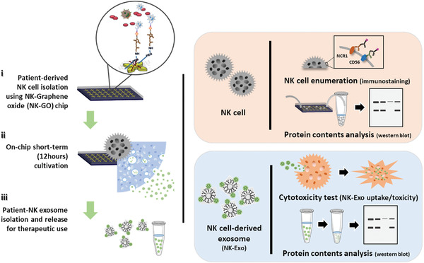
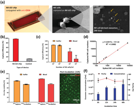
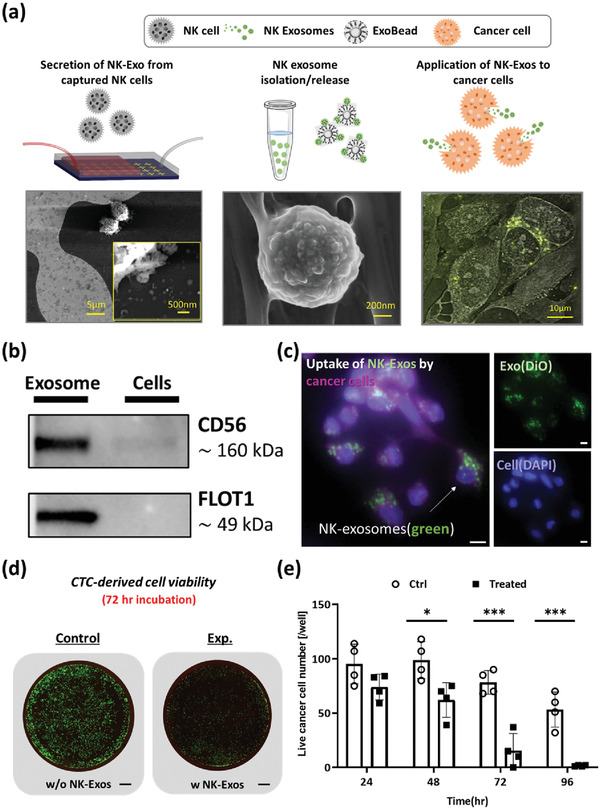
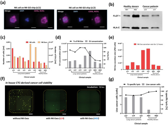
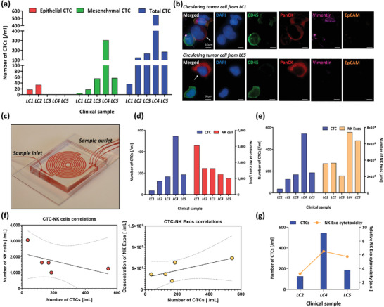
As the recognition between natural killer (NK) cells and cancer cells does not require antigen presentation, NK cells are being actively studied for use in adoptive cell therapies in the rapidly evolving armamentarium of cancer immunotherapy. In addition to utilizing NK cells, recent studies have shown that exosomes derived from NK cells also exhibit antitumor properties. Furthermore, these NK cell-derived exosomes exhibit higher stability, greater modification potentials and less immunogenicity compared to NK cells. Therefore, technologies that allow highly sensitive and specific isolation of NK cells and NK cell-derived exosomes can enable personalized NK-mediated cancer therapeutics in the future. Here, a novel microfluidic system to collect patient-specific NK cells and on-chip biogenesis of NK-exosomes is proposed. In a small cohort of non-small cell lung cancer (NSCLC) patients, both NK cells and circulating tumor cells (CTCs) were isolated, and it is found NSCLC patients have high numbers of NK and NK-exosomes compared with healthy donors, and these concentrations show a trend of positive and negative correlations with bloodborne CTC numbers, respectively. It is further demonstrated that the NK-exosomes harvested from NK-graphene oxide chip exhibit cytotoxic effect on CTCs. This versatile system is expected to be used for patient-specific NK-based immunotherapies along with CTCs for potential prognostic/diagnostic applications.

Keywords: cancer immunotherapy; circulating tumor cells; exosome biogenesis; microfluidics; natural killer cells.


Conflict of interest statement

S.N. is one of the named inventors on a patent for Microfluidic Labyrinth Technology granted to the University of Michigan. S.N. is also the co‐founder of Labyrinth Biotech Inc. The funders and the company had no role in the design of the study; in the collection, analyses, or interpretation of data; in the writing of the manuscript; or in the decision to publish the results.

Figures

Figure 1
Figure 1
Schematic diagram of the microfluidic technology approach for on‐chip natural killer (NK) cell isolation, in situ NK cell‐derived exosome biogenesis, and recovery for potential therapeutic use of NK exosomes.
Figure 2
Figure 2
On‐chip NK cell isolation and in situ exosome biogenesis and harvesting from the isolated NK cells: a) in situ exosome biogenesis from isolated NK cells on NK‐GO chip; b) NK cell capturing performance of NK‐GO chip compared to control device without antibody conjugation; c) capture efficiency of the NK‐GO chip for different concentrations of NK cells spiked in buffer and blood samples; d) captured NK cells depending on spiked NK cells in buffer solution. The red dashed line is a linear fit to the data; e) viability of the isolated NK cells at different time points and conditions (left) and live/dead staining of isolated NK cells on chip after 12 h incubation; f) exosome secretion from NK cells depending on incubation times.
Figure 3
Figure 3
ExoBead‐based NK exosome isolation and release for therapeutic use: a) scanning electron microscope image of the isolated exosomal vesicles on ExoBeads with supernatant from NK‐GO chip after 12 h incubation; b) concentration and purity of exosomal vesicles recovered from NK‐92MI culture supernatant under three different conditions using ExoBeads (ExoB) and control beads (ConB) nonconjugated with antibodies; c) capture and release performance of ExoBeads depending on amounts of beads for identical volume of NK‐GO chip supernatant; d) capture and recovery performance of ExoBeads depending on amounts of d‐biotinylated anti‐CD63 during antibody conjugation; e) release and purity performance comparison between two different incubation times with biotin solution for exosome release.
Figure 4
Figure 4
Cytotoxicity of NK cell‐derived exosomes: a) NK cells (left) and NK exosomes (center) recovered from current platform and theranostic use of NK exosomes with cancer cells (right); b) western blots showing the positive expression of CD56 and FLOT1 in exosome lysate using a NK‐92MI spike in buffer sample for on‐chip exosome biogenesis. The cell lysate from the same device also show positive expression for CD56; c) uptake of NK‐92MI exosomes by in‐house CTC‐derived cell line. Exosomes are fluorescently labeled in green (FITC) channel, and cell membrane is labeled in violet (Cy5) (Scale bar = 10 µm); d) cytotoxicity comparison between control well and NK exosome treated well at 72 h (Scale bar = 800 µm); e) in vitro cytotoxicity experiment using exosomes derived from NK‐92MI. Live cells were quantified through a live/dead assay that was performed 24, 48, and 72 h after treating cancer cells with or without NK exosomes. Unpaired t‐tests (two‐tailed) were used to compare the differences between live cell count between NK exosome treated (n = 4) versus control (n = 4). Asterisks denote one of three levels of statistical significance (*p ≤ 0.05; **p ≤ 0.01; ***p ≤ 0.001).
Figure 5
Figure 5
Analysis of clinical samples from NSCLC patients using NK‐GO microfluidic platform: a) immunofluorescence image examples of CD56/NCR1 + NK cells captured on NK‐GO chip (Scale bar = 20 µm); b) western blot analysis for showing the positive expression of FLOT1 and HLA‐C in exosomes from clinical samples; c) profiling in quantity of NK cells and NK cell‐derived exosomes among different patients and healthy individuals observed after 12 h on‐chip incubation; d) total extracellular vesicle concentration and percentage of exosomes among patient samples and healthy control samples; e) biogenesis of exosomes quantified as secretion rate of exosomes per captured NK cells for 12 h; f) Live cancer cell number after 72 h incubation and percentage of specific lysis between samples from cancer patients and healthy donors (Scale bar = 20 µm); g) cytotoxicity of clinical sample driven NK‐Exos to CTC‐derived cells after 72 h incubation.
Figure 6
Figure 6
Profiling of circulating tumor cell (CTC) populations in non‐small cell lung cancer (NSCLC) patients and correlation with NK cells and NK cell‐derived exosomes: a) profiling in quantity of epithelial, mesenchymal, and total CTCs between NSCLC patients; b) representative images of CTCs recovered from the patients; c) image of label‐free CTC isolation platform; d) comparison between total CTC and NK cell number; e) comparison between total CTC and NK exosome concentration; f) correlation between CTC‐NK cell (left, r = −0.580, P‐value = 0.305) or CTC‐NK‐Exo (right, r = 0.732, P‐value = 0.159); g) correlation between relative total cytotoxicity of NK‐Exos and total CTC number in samples.

Similar articles

Cited by

References

    1. Davis I. D., Immunol. Cell Biol. 2000, 78, 179. - PubMed
    1. June C. H., J. Clin. Invest. 2007, 117, 1204. - PMC - PubMed
    1. Dine J., Gordon R., Shames Y., Kasler M. K., Barton‐Burke M., Asia Pac. J. Oncol. Nurs. 2017, 4, 127. - PMC - PubMed
    1. Barbee M. S., Ogunniyi A., Horvat T. Z., Dang T. O., Ann. Pharmacother. 2015, 49, 907. - PubMed
    1. Gattinoni L., Semin. Immunol. 2016, 278, 1. - PMC - PubMed