

The changing face of central chondrosarcoma of bone. One UK-based orthopaedic oncology unit's experience of 33 years referrals
- PMID: 33747783
- PMCID: PMC7972956
- DOI: 10.1016/j.jcot.2021.02.017
The changing face of central chondrosarcoma of bone. One UK-based orthopaedic oncology unit's experience of 33 years referrals
Abstract
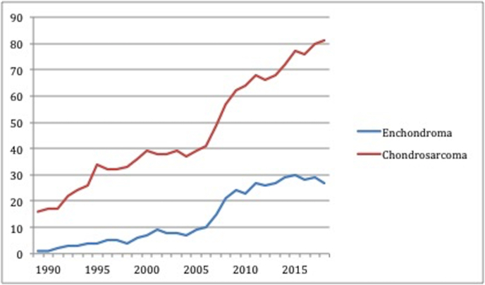
Aim: To ascertain the changing incidence over time of the three commonest primary sarcomas of bone. Data obtained with particular reference to central chondrosarcoma from the annual referral rate to a large UK-based specialist orthopaedic oncology unit. To discuss how the "barnyard pen" analogy of cancers previously applied to certain commoner cancers can also be applicable to central chondrosarcoma (CS) of bone.
Materials and methods: A retrospective review was conducted of a computerised database identifying all central cartilage tumours (CCT) of bone, including enchondroma and CS subtypes, between 1985 and 2018. These were compared with the referrals of the other two commonest primary sarcomas of bone, osteosarcoma and Ewing sarcoma.
Results: There was a total of 1507 CS showing a 68% overall increase in annual referral rate/incidence over the study period. 68% cases were the borderline malignant lesions now known as atypical cartilaginous tumour (ACT). The annual referral rate/incidence of this entity increased by 194% over the 30 years. Whereas, the annual referral rate/incidence for osteosarcoma and Ewing sarcoma was static for the past 20 years.
Conclusion: The annual incidence of central CS of bone showed a marked increase over the 33-year period as compared with both osteosarcoma and Ewing sarcoma. This is especially in the ACT category and is thought to be due to the increased provision of MRI scanning flagging up a rise in incidental findings. The spectrum of CCTs from benign to highly malignant elegantly fits the "barn yard" pen analogy and could prove useful as an explanatory tool for patients and clinicians unfamiliar with these diseases.
Keywords: Atypical cartilage tumour (ACT); Chondrosarcoma; Enchondroma.
© 2021 Delhi Orthopedic Association. All rights reserved.
Figures


References
-
- Hogendoorn P.C.W., Bovee J.V.M.G., Nielsen G.P. Chondrosarcoma (grades I-III) In: Fletcher C.D.M., Bridge J.A., Hogendoorn P.C.W., editors. WHO Classification of Tumours of Soft Tissue and Bone. International Agency for Research on Cancer; Lyon: 2013. pp. 264–268.
-
- Skeletal Lesions Interobserver Correlation among Expert Diagnosticians (SLICED) Study Group Reliability of histopathologic and radiologic grading of cartilaginous neoplasms of long bones. J Bone Joint Surg. 2007;89A:2113–2123. - PubMed
-
- Eefting D., Schrage Y.M., Geirnaerdt M.J. EuroBoneNet consortium. Assessment of interobserver variability and histologic parameters to improve reliability in classification and grading central cartilaginous tumors. Am J Surg Pathol. 2009;33:50–57. - PubMed
-
- Crim J., Schmidt R., Layfield L. Can imaging criteria distinguish enchondroma from grade I chondrosarcoma? Eur J Radiol. 2015;84:2222–2230. - PubMed
-
- Fritz B., Muller D.A., Sutter R. Magnetic resonance imaging-based grading of cartilaginous bone tumors: added value of quantitative texture analysis. Invest Radiol. 2018;53:663–672. - PubMed
LinkOut - more resources
Full Text Sources
Other Literature Sources
Miscellaneous