

Comparison of healing patterns of different side-cut angulations after FS-LASIK
- PMID: 33747821
- PMCID: PMC7930535
- DOI: 10.18240/ijo.2021.03.16
Comparison of healing patterns of different side-cut angulations after FS-LASIK
Abstract
Aim: To investigate and evaluate healing patterns around flaps made with different side-cut angulations after femtosecond laser in situ keratomileusis (FS-LASIK).
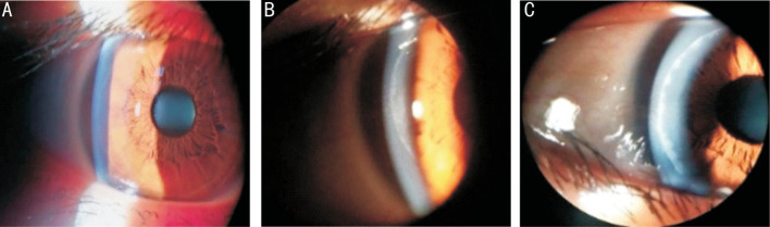
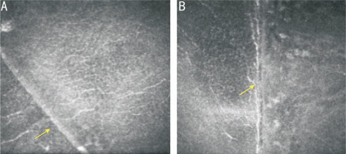
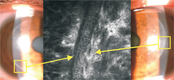
Methods: Thirty-four patients (68 eyes) received a 90° side-cut (n=34) or a 120° side-cut flaps (n=34) made with a femtosecond laser. One day, 1wk, 1 and 3mo postoperatively, side-cut scar was evaluated under slit-lamp photography according to a new grading system (Grade 0=transparent scar, 1=faint healing opacity, and 2=evident healing opacity). In vivo corneal confocal microscopy and anterior segment optical coherence tomography (AS-OCT) were used to observe wound-healing patterns around flap margin in the two groups. Sirius Scheimpflug Analyzer was also used to analyze higher order aberrations 3mo after surgery.
Results: There were no significant differences in flap wound-healing patterns at each follow up between the two groups (P>0.05). Three months after surgery, the flap edge scar classified as Grade 0 had excellent apposition and rapid nerve regeneration. At 3 mm and 5 mm pupil diameters, there were significant differences in trefoil aberrations between the two groups (P<0.05), but no statistically significant differences were found in total higher order aberrations (HOAs), spherical aberrations or coma in any of the pupil size conditions (P>0.05).
Conclusion: Flap edge scars classified as Grade 0 have excellent apposition and rapid nerve regeneration, and 120° side-cut angle flaps induce less trefoil aberrations after FS-LASIK.
Keywords: confocal microscopy; femtosecond laser; healing; scar; side-cut.
International Journal of Ophthalmology Press.
Figures

Similar articles
-
Comparison of Laser In Situ Keratomileusis Flap Morphology and Predictability by WaveLight FS200 Femtosecond Laser and Moria Microkeratome: An Anterior Segment Optical Coherence Tomography Study.Korean J Ophthalmol. 2019 Apr;33(2):113-121. doi: 10.3341/kjo.2018.0035. Korean J Ophthalmol. 2019. PMID: 30977320 Free PMC article.
-
[Comparison of the effects of different side-cut angles on corneal biomechanical properties after femtosecond laser assisted-laser in situ keratomileusis].Zhonghua Yan Ke Za Zhi. 2017 Jan 11;53(1):23-32. doi: 10.3760/cma.j.issn.0412-4081.2017.01.006. Zhonghua Yan Ke Za Zhi. 2017. PMID: 28162196 Chinese.
-
Three-year results of small incision lenticule extraction and wavefront-guided femtosecond laser-assisted laser in situ keratomileusis for correction of high myopia and myopic astigmatism.Int J Ophthalmol. 2018 Mar 18;11(3):470-477. doi: 10.18240/ijo.2018.03.18. eCollection 2018. Int J Ophthalmol. 2018. PMID: 29600182 Free PMC article.
-
Comparison of the femtosecond laser and mechanical microkeratome for flap cutting in LASIK.Int J Ophthalmol. 2015 Aug 18;8(4):784-90. doi: 10.3980/j.issn.2222-3959.2015.04.25. eCollection 2015. Int J Ophthalmol. 2015. PMID: 26309880 Free PMC article.
-
Verification and measurement of the side-cut angle of corneal flap in patients undergoing LASIK surgery using FS 200 kHz femtosecond laser system versus conventional mechanical microkeratome.Clin Ophthalmol. 2019 Jun 10;13:985-992. doi: 10.2147/OPTH.S201150. eCollection 2019. Clin Ophthalmol. 2019. PMID: 31354232 Free PMC article.
References
-
- Vaddavalli PK, Yoo SH. Femtosecond laser in situ keratomileusis flap configurations. Curr Opin Ophthalmol. 2011;22(4):245–250. - PubMed
-
- Slade S, Ignacio T, Spector S. Evaluation of a multifunctional femtosecond laser for the creation of laser in situ keratomileusis flaps. J Cataract Refract Surg. 2018;44(3):280–286. - PubMed
-
- Knox Cartwright NE, Tyrer JR, Jaycock PD, Marshall J. Effects of variation in depth and side cut angulations in LASIK and thin-flap LASIK using a femtosecond laser: a biomechanical study. J Refract Surg Thorofare N J. 2012;28(6):419–425. - PubMed
-
- Knorz MC, Vossmerbaeumer U. Comparison of flap adhesion strength using the Amadeus microkeratome and the IntraLase iFS femtosecond laser in rabbits. J Refract Surg. 2008;24(9):875–878. - PubMed
LinkOut - more resources
Full Text Sources
Other Literature Sources
Research Materials