. 2021 Mar 3:11:597503.
doi: 10.3389/fonc.2021.597503. eCollection 2021.

Anti-Tumoral and Anti-Angiogenic Effects of Low-Diluted Phenacetinum on Melanoma

Affiliations

Anti-Tumoral and Anti-Angiogenic Effects of Low-Diluted Phenacetinum on Melanoma

Camille Fuselier et al. Front Oncol. .

Abstract

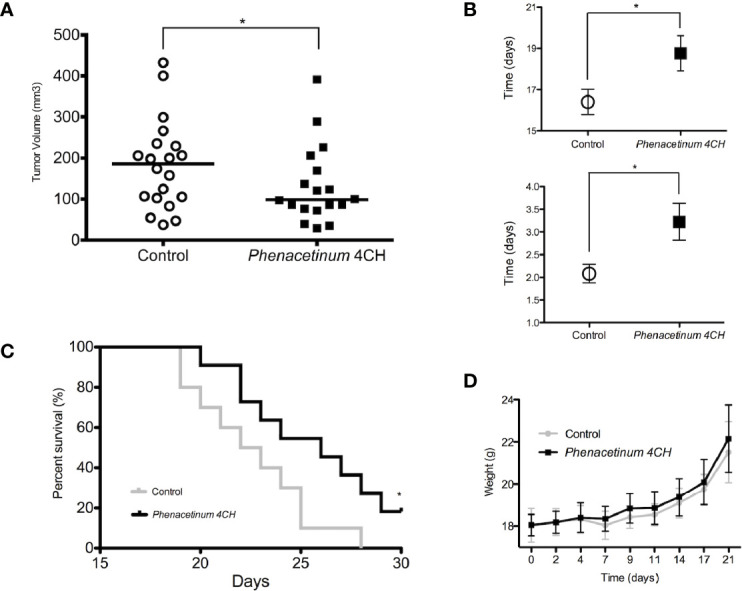
Melanoma is the most aggressive form of skin cancer and the most rapidly expanding cancer in terms of worldwide incidence. If primary cutaneous melanoma is mostly treated with a curative wide local excision, malignant melanoma has a poor prognosis and needs other therapeutic approaches. Angiogenesis is a normal physiological process essential in growth and development, but it also plays a crucial role in crossing from benign to advanced state in cancer. In melanoma progression, angiogenesis is widely involved during the vertical growth phase. Currently, no anti-angiogenic agents are efficient on their own, and combination of treatments will probably be the key to success. In the past, phenacetin was used as an analgesic to relieve pain, causing side effects at large dose and tumor-inducing in humans and animals. By contrast, Phenacetinum low-dilution is often used in skin febrile exanthema, patches profusely scattered on limbs, headache, or flushed face without side effects. Herein are described the in vitro, in vivo, and ex vivo anti-angiogenic and anti-tumoral potentials of Phenacetinum low-dilution in a B16F1 tumor model and endothelial cells. We demonstrate that low-diluted Phenacetinum inhibits in vivo tumor growth and tumor vascularization and thus increases the survival time of B16F1 melanoma induced-C57BL/6 mice. Moreover, Phenacetinum modulates the lung metastasis in a B16F10 induced model. Ex vivo and in vitro, we evidence that low-diluted Phenacetinum inhibits the migration and the recruitment of endothelial cells and leads to an imbalance in the pro-tumoral macrophages and to a structural malformation of the vascular network. All together these results demonstrate highly hopeful anti-tumoral, anti-metastatic, and anti-angiogenic effects of Phenacetinum low-dilution on melanoma. Continued studies are needed to preclinically validate Phenacetinum low-dilution as a complementary or therapeutic strategy for melanoma treatment.

Keywords: angiogenesis; cancer; homeopathy; in vivo; melanoma; metastasis; phenacetin; tumor-associated macrophages.


Conflict of interest statement

The authors declare that the research was conducted in the absence of any commercial or financial relationships that could be construed as a potential conflict of interest.

Figures

Figure 1
Figure 1
Effect of Phenacetinum 4CH on tumor progression, body weight, and survival time 2.5 x 10⁵ melanoma B16F1 cells were subcutaneously injected into the left side of C57BL/6 mice. Control (open circle) or Phenacetinum 4CH (filled box) (100 µl) were intraperitoneally injected each day. (A) Tumor volume was measured with a caliper as described in Materials and Methods. Graph represents tumor volume measurements at day 14 after melanoma cells injection. Horizontal lines, median (n = 20, Mann-Whitney test). (B) Time to reach a tumor volume of 300 mm3 (1B high) and doubling tumor volume time (1B low) during the exponential growth phase (days 14 to 17). (C) The survival rates of C57Bl/6 mice with B16F1 melanoma xenografts treated with Control (grey line) or Phenacetinum 4CH (black line) were recorded and shown as Kaplan-Meir curves. (D) Mouse mean weight evolution for control (grey line) and Phenacetinum 4CH (black line) groups. (*p < 0.05).
Figure 2
Figure 2
Effect of Phenacetinum 4CH on in vivo tumor vascularization. (A, B) are representative tridimensional reconstructions of tumors at day 14 after µCT reconstruction. Melanoma tumors were segmented, and the associated vascular networks were traced using filament editor of Amira 6.5 software. Color-coded representation of tumor-associated blood vessel network depended on structure thickness (Amira 6.5 software). Thin structures are represented in blue, which changes to green and yellow when the vessel diameter increases. (C) is quantification of tumor-associated blood vessel mean length, volume, and radius using Amira 6.5 software. Data are expressed as the mean +/- SEM (*p < 0.05).
Figure 3
Figure 3
Inhibition of Matrigel® plug angiogenesis by Phenacetinum 4CH. FGF- and VEGF-induced Matrigel® plug angiogenesis assay was performed on the back on C57Bl/6 mice. Mice were perfused, injected with 0.1 mmol/kg of Galodidium, and DCE-MRI analyzed. (A) Matrigel® plug’ ROI intensity of control mice during perfusion. (B) Matrigel® plug’ ROI intensity of Phenacetinum 4CH mice during perfusion. (C) Pool of control (black) and Phenacetinum 4CH (gray) ROI intensity curves and wash-in rate tangents.
Figure 4
Figure 4
Effect of Phenacetinum 4CH on ex vivo necrosis, CD31, CD163, and CD68 expression. Photographs 4A and 4C show representative tumor sections from control or Phenacetinum 4CH treated mice. (A) HPS staining of tumor sections (left panel: control, right panel: Phenacetinum 4CH) showing tumor necrosis (area delimited by continuous line). (B) Semi-quantitative analysis with Image J software of necrosis area (% of total surface). (C) IHC staining in control tumor (left column) and Phenacetinum-treated tumor (right column) using anti-CD31, CD163, and CD68 antibodies. (D) Semi-quantitative with image J software analysis of CD31 staining (% of total surface) and expression of CD163+ and CD68+ cells (number/10 High-Power Fields). Data are expressed as the mean +/- SEM (*p < 0.05, **p < 0.01).
Figure 5
Figure 5
Effect of Phenacetinum 4CH on ex vivo aortic rings explants. Aorta were ex vivo seeded on Matrigel®. Phase contrast photos were taken at day 3, 5, and 7. (A) Representative photograph and modelling of control aortic assays using Image J software. (B) Representative photograph and modelling of Phenacetinum 4CH aortic assays using Image J software. (C–F) Semi-quantification at days 3, 5, and 7 of aortic rings network length (C), surface (D), junctions (E), and ending (F). (*p < 0.05, **p < 0.01).
Figure 6
Figure 6
Effect of Phenacetinum 4CH on in vitro HUVEC tubulogenesis, migration, and proliferation. For tubulogenesis, HUVEC cells were seeded on Matrigel®. Phase contrast overview photos were taken after 6 h. For 3D migration, HUVEC cells were seeded on transwell coated with Collagen I and migrated cells on the lower membrane counted after 18 h. (A) Representative photo of control HUVEC tubulogenesis on Matrigel®. (B) Representative photo of Phenacetinum 4CH HUVEC tubulogenesis on Matrigel®. (C) Quantification of isolated segments. (D) Quantification of isolated branched length. (E) Quantification of HUVEC migrated cells with B16F1 (clear boxes) or B16F10 (gray boxes) conditioned media as chemoattractant. (F) Quantification of HUVEC proliferation. (ns, not significant, *p < 0.05, **p < 0.01, ***p < 0.001).
Figure 7
Figure 7
Effect of Phenacetinum 4CH on melanoma metastatic dissemination. 1 x 10⁵ melanoma B16F10 cells were injected in the lateral vein of C57BL/6 mice. Control (open circle) or Phenacetinum 4CH (filled box) (100 µl) were intraperitoneally injected each day. (A) Metastatic surface ratio (metastases/health lungs) at day 21 (median, n=10, Mann-Whitney test, **p < 0.01). (B) representative tridimensional reconstructions of mice at day 14 after µCT reconstruction. Metastases were segmented using Amira 6.5 software. Metastases are represented in blue. (C) T2 weighted images of the lungs from control (upper panel) and Phenacetinum 4CH-treated (lower panel) mice. Metastases appear as white opaque hyper-intense regions. (D) HPS staining of lungs section (left panel: control, right panel: Phenacetinum 4CH). Arrow identified metastatic foci.

Similar articles

Cited by

References

    1. Miller AJ, Mihm MC. Melanoma. N Engl J Med (2006) 355:51–65. 10.1056/NEJMra052166 - DOI - PubMed
    1. Torre LA, Bray F, Siegel RL, Ferlay J, Lortet-Tieulent J, Jemal A. Global cancer statistics, 2012. CA Cancer J Clin (2015) 65:87–108. 10.3322/caac.21262 - DOI - PubMed
    1. Schadendorf D, van Akkooi ACJ, Berking C, Griewank KG, Gutzmer R, Hauschild A, et al. . Melanoma. Lancet Lond Engl (2018) 392:971–84. 10.1016/S0140-6736(18)31559-9 - DOI - PubMed
    1. Dimitriou F, Krattinger R, Ramelyte E, Barysch MJ, Micaletto S, Dummer R, et al. . The World of Melanoma: Epidemiologic, Genetic, and Anatomic Differences of Melanoma Across the Globe. Curr Oncol Rep (2018) 20:87. 10.1007/s11912-018-0732-8 - DOI - PubMed
    1. Shain AH, Bastian BC. From melanocytes to melanomas. Nat Rev Cancer (2016) 16:345–58. 10.1038/nrc.2016.37 - DOI - PubMed

LinkOut - more resources