. 2021 Mar 3:9:642866.
doi: 10.3389/fbioe.2021.642866. eCollection 2021.

Three-Dimensional Quantification of Collagen Microstructure During Tensile Mechanical Loading of Skin

Affiliations

Three-Dimensional Quantification of Collagen Microstructure During Tensile Mechanical Loading of Skin

Alan E Woessner et al. Front Bioeng Biotechnol. .

Abstract

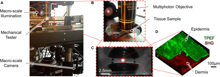
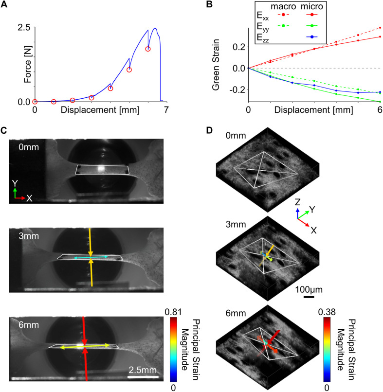
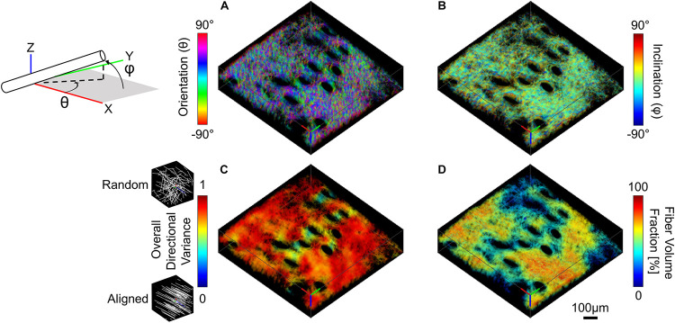
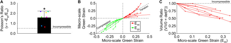
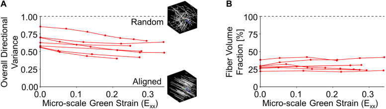
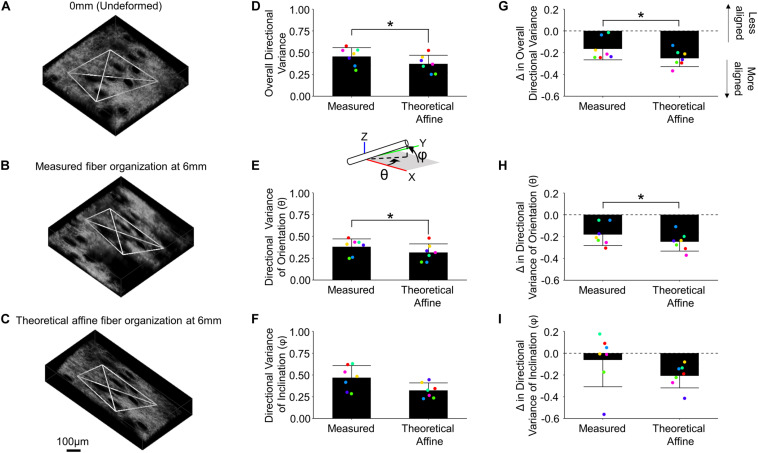
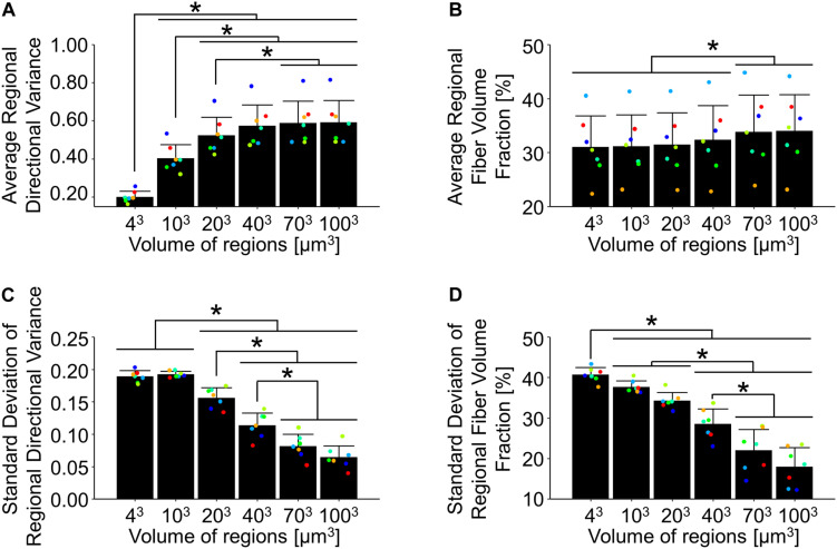
Skin is a heterogeneous tissue that can undergo substantial structural and functional changes with age, disease, or following injury. Understanding how these changes impact the mechanical properties of skin requires three-dimensional (3D) quantification of the tissue microstructure and its kinematics. The goal of this study was to quantify these structure-function relationships via second harmonic generation (SHG) microscopy of mouse skin under tensile mechanical loading. Tissue deformation at the macro- and micro-scale was quantified, and a substantial decrease in tissue volume and a large Poisson's ratio was detected with stretch, indicating the skin differs substantially from the hyperelastic material models historically used to explain its behavior. Additionally, the relative amount of measured strain did not significantly change between length scales, suggesting that the collagen fiber network is uniformly distributing applied strains. Analysis of undeformed collagen fiber organization and volume fraction revealed a length scale dependency for both metrics. 3D analysis of SHG volumes also showed that collagen fiber alignment increased in the direction of stretch, but fiber volume fraction did not change. Interestingly, 3D fiber kinematics was found to have a non-affine relationship with tissue deformation, and an affine transformation of the micro-scale fiber network overestimates the amount of fiber realignment. This result, along with the other outcomes, highlights the importance of accurate, scale-matched 3D experimental measurements when developing multi-scale models of skin mechanical function.

Keywords: collagen; kinematics; microstructure; multiscale; second harmonic generation; skin; structure-function relationship.


Conflict of interest statement

The authors declare that the research was conducted in the absence of any commercial or financial relationships that could be construed as a potential conflict of interest.

Figures

FIGURE 1
FIGURE 1
A mechanical tensile testing device integrated with a multi-scale imaging system (A,B) was used to collect macro-scale 2D images [(C); scale bar represents 2.5 mm] and micro-scale 3D MPM image volumes [(D); scale bar represents 100 μm]. To highlight the difference in scale between the macro- and micro-scale ROIs, the red dashed square in the center region of panel (C) represents the relative size of the ROI collected in panel (D). At the micro-scale, both SHG and TPEF image volumes were collected, and the pseudo-colored TPEF volume shown in panel (D) is cutout to highlight the layers of the skin.
FIGURE 2
FIGURE 2
Tissue kinematics were quantified at multiple length scales during incremental stretching of skin in the x-direction. (A) To enable the collection of high-resolution micro-scale SHG image volumes, the skin was stretched in 1 mm increments (red circles) until mechanical failure. (B) Tissue kinematics were restricted to 2D at the macro-scale but were quantified in 3D at the micro-scale. (C) Macro-scale tissue deformation measured from fiduciary markers showed a substantial compressive principal strain orthogonal to the direction of stretch (scale bar represents 2.5 mm). (D) 3D micro-scale tissue kinematics measured from SHG features indicated compressive principal strains were occurring in both directions orthogonal to the loading direction (scale bar represents 100 μm). For panels (C,D), white polygons indicate tracked features, and arrows represent the direction and magnitude of principal strains.
FIGURE 3
FIGURE 3
SHG image volumes were used to compute a pixel-wise map of 3D micro-scale orientation (A) and inclination (B). The two 3D orientation and inclination maps and collagen-positive mask were then convolved with a range of cubic averaging kernels varying in size to determine how regional measurements of directional variance (C) and fiber volume fraction (D) change as a function of the length scale over which they are measured (convolution with 40 μm3 kernel shown here, scale bar represents 100 μm).
FIGURE 4
FIGURE 4
Tissue kinematics measured at multiple length scales were found to experience relatively similar magnitudes of strain and exhibit compressible material behaviors. (A) At the macro-scale, tissue exhibited a very large Poisson’s ratio that was significantly different than expected from an incompressible material. (B) Macro- and micro-scale Green strain for all samples indicates similar tissue deformation across length-scales. (C) Tissue underwent a significant decrease in micro-scale volume during stretch.
FIGURE 5
FIGURE 5
Collagen fiber directional variance decreased during stretch (A), indicating increased alignment, but fiber volume fraction did not significantly change (B).
FIGURE 6
FIGURE 6
Collagen fiber kinematics are not affine with the local skin deformation. Micro-scale image volumes were compared among the undeformed configuration (A), actual tissue response at peak strain (B) and the theoretical affine transformation (C) based on the measured deformation gradient. The white tetrahedron in panels (A–C) indicates the position of 3D image features that were tracked during deformation (scale bar represents 100 μm). The computed 3D directional variance (D) as well as the measured directional variance for individual 3D orientation and inclination components [θ and φ, panels (E,F), respectively] were found to be higher than the theoretical affine transformation. Similarly, the change in overall directional variance (G) and the 3D orientation and inclination components (H,I) indicate the measured fiber re-alignment in the direction of loading is less than the theoretical affine response. Points are colored to indicate paired comparisons among groups. *represents p < 0.05.
FIGURE 7
FIGURE 7
In the undeformed configuration, regional averages of directional variance (A) and fiber volume fraction (B) increased as a function of the length scale over which they were measured. Alternatively, the average regional standard deviations of directional variance (C) and fiber volume fraction (D) decreased at larger length-scales, indicating the importance of length-scale when implementing measurements into computational models. Points are colored to indicate paired comparisons among groups. *represents p < 0.05.

Similar articles

Cited by

References

    1. Alberto R., Draicchio F., Varrecchia T., Silvetti A., Iavicoli S. (2018). Wearable monitoring devices for biomechanical risk assessment at work: current status and future challenges-a systematic review. Int. J. Environ. Res. Public Health 15:2001. 10.3390/ijerph15092001 - DOI - PMC - PubMed
    1. Aubert L., Anthoine P., Rigal J. D., Leveque J. L. (1985). An in vivo assessment of the biomechanical properties of human skin modifications under the influence of cosmetic products. Int. J. Cosmet. Sci. 7 51–59. 10.1111/j.1467-2494.1985.tb00396.x - DOI - PubMed
    1. Ban E., Wang H., Franklin J. M., Liphardt J. T., Janmey P. A., Shenoy V. B. (2019). Strong triaxial coupling and anomalous Poisson effect in collagen networks. Proc. Natl. Acad. Sci. U.S.A. 116 6790–6799. 10.1073/pnas.1815659116 - DOI - PMC - PubMed
    1. Bancelin S., Aime C., Gusachenko I., Kowalczuk L., Latour G., Coradin T., et al. (2014). Determination of collagen fibril size via absolute measurements of second-harmonic generation signals. Nat. Commun. 5:4920. 10.1038/ncomms5920 - DOI - PubMed
    1. Bancelin S., Lynch B., Bonod-Bidaud C., Ducourthial G., Psilodimitrakopoulos S., Dokladal P., et al. (2015). Ex vivo multiscale quantitation of skin biomechanics in wild-type and genetically-modified mice using multiphoton microscopy. Sci. Rep. 5:17635. 10.1038/srep17635 - DOI - PMC - PubMed

LinkOut - more resources