Review
. 2021 Mar 5:9:623734.
doi: 10.3389/fcell.2021.623734. eCollection 2021.

On the Wrong Track: Alterations of Ciliary Transport in Inherited Retinal Dystrophies

Affiliations
Review

On the Wrong Track: Alterations of Ciliary Transport in Inherited Retinal Dystrophies

Laura Sánchez-Bellver et al. Front Cell Dev Biol. .

Abstract

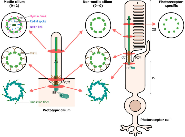
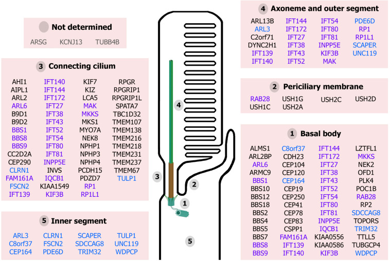
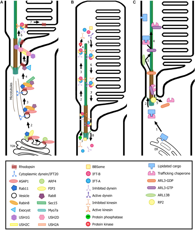
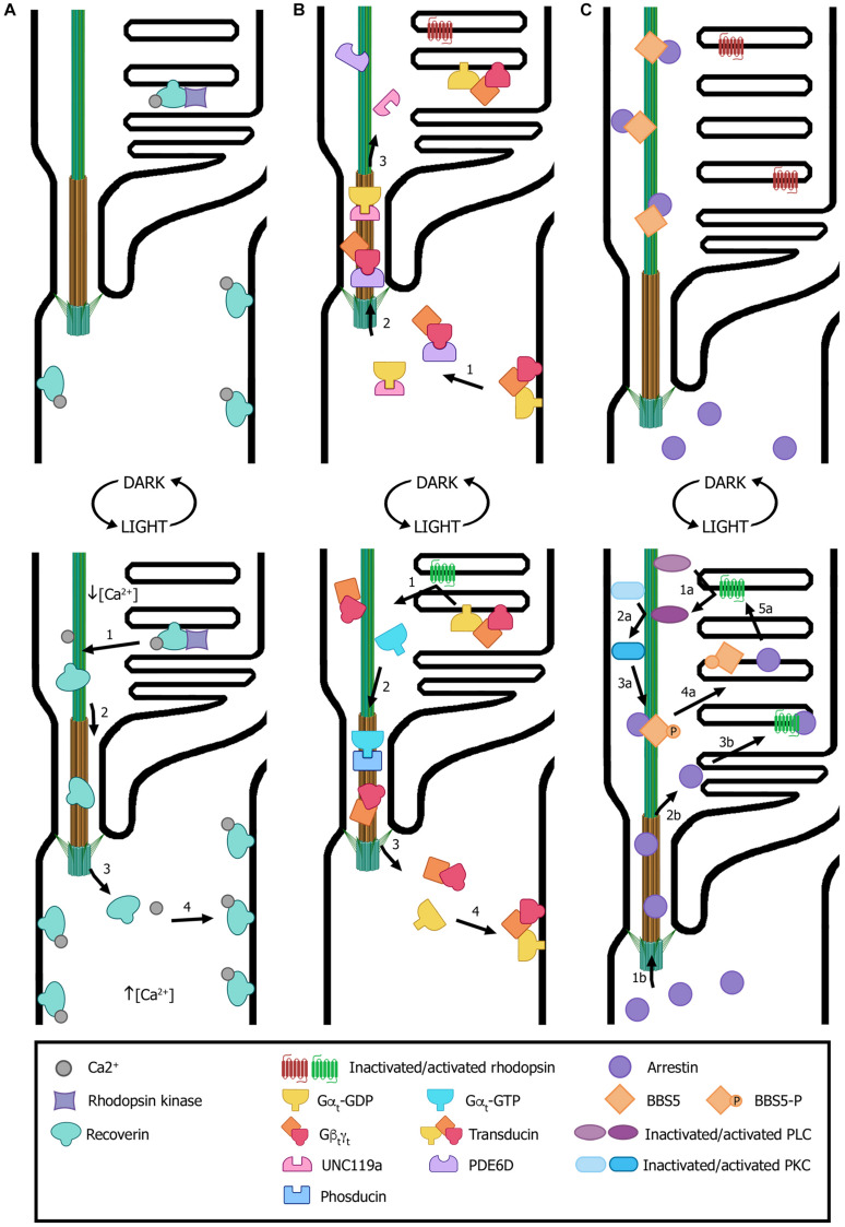
Ciliopathies are a group of heterogeneous inherited disorders associated with dysfunction of the cilium, a ubiquitous microtubule-based organelle involved in a broad range of cellular functions. Most ciliopathies are syndromic, since several organs whose cells produce a cilium, such as the retina, cochlea or kidney, are affected by mutations in ciliary-related genes. In the retina, photoreceptor cells present a highly specialized neurosensory cilium, the outer segment, stacked with membranous disks where photoreception and phototransduction occurs. The daily renewal of the more distal disks is a unique characteristic of photoreceptor outer segments, resulting in an elevated protein demand. All components necessary for outer segment formation, maintenance and function have to be transported from the photoreceptor inner segment, where synthesis occurs, to the cilium. Therefore, efficient transport of selected proteins is critical for photoreceptor ciliogenesis and function, and any alteration in either cargo delivery to the cilium or intraciliary trafficking compromises photoreceptor survival and leads to retinal degeneration. To date, mutations in more than 100 ciliary genes have been associated with retinal dystrophies, accounting for almost 25% of these inherited rare diseases. Interestingly, not all mutations in ciliary genes that cause retinal degeneration are also involved in pleiotropic pathologies in other ciliated organs. Depending on the mutation, the same gene can cause syndromic or non-syndromic retinopathies, thus emphasizing the highly refined specialization of the photoreceptor neurosensory cilia, and raising the possibility of photoreceptor-specific molecular mechanisms underlying common ciliary functions such as ciliary transport. In this review, we will focus on ciliary transport in photoreceptor cells and discuss the molecular complexity underpinning retinal ciliopathies, with a special emphasis on ciliary genes that, when mutated, cause either syndromic or non-syndromic retinal ciliopathies.

Keywords: ciliary transport; ciliopathy; inherited retinal dystrophies; intraflagellar transport; photoreceptor; sensory cilium.


Conflict of interest statement

The authors declare that the research was conducted in the absence of any commercial or financial relationships that could be construed as a potential conflict of interest.

Figures

FIGURE 1
FIGURE 1
Structure of motile, non-motile, and photoreceptor cilia. The left diagram depicts a prototypic cilium with its main compartments: the axoneme (Ax), transition zone (TZ) with its Y-links, periciliary membrane (PCM) and the basal body (BB). The right diagram depicts a rod photoreceptor, which in addition, displays photoreceptor-specific cellular structures: the outer segment (OS), comprising the membranous disks (D) and the axoneme; the connecting cilium (CC), analogous to the transition zone in primary cilia; and the inner segment (IS), which contains the basal body and most cellular organelles. Cross-sections through the ciliary compartments of both motile and non-motile cilia are shown: the basal body triplet microtubules with their transition fibers, shared by motile cilia, non-motile cilia and photoreceptor primary cilia; the transition zone of motile and non-motile cilia and the analogous connecting cilium in photoreceptors; the axonemal microtubule doublets, decorated with dynein arms, radial spokes and nexin links in motile cilia; and finally, the axonemal microtubule singlets characteristic of photoreceptor primary cilia. (Figure created with BioRender.com).
FIGURE 2
FIGURE 2
Proteins associated with retinal ciliopathies and their reported localization at the photoreceptor structures. Ciliary proteins can localize to unique ciliary subcompartments (names in black), several ciliary subcompartments (highlighted in purple) or other cellular compartments in addition to the cilium (in blue). The localization of ARSG, KCNJ13, and TUBB4B has not been determined yet (in gray). (Figure created with BioRender.com).
FIGURE 3
FIGURE 3
Transport of ciliary proteins in photoreceptor cells. (A) Rhodopsin is transported from the trans-Golgi network (TGN) to the rod outer segment by a conventional secretory pathway regulated by small GTPases. Many ciliary protein complexes are involved in this process, performing different roles in trafficking that enable rhodopsin docking and transport through the connecting cilium to the ciliary membrane. Note the location of the USH protein complex formed by USH1G, USH2D, USH2A, and USH2C subunits. (B) The proposed model for the cycle of the IFT complexes in photoreceptors is based on flagella and primary cilium as well as cryo-EM structural models (Liang et al., 2014; Roberts, 2018; Toropova et al., 2019). IFT-B trains (responsible for anterograde trafficking) are assembled at the base of the cilium and move along the outer segment axoneme to the tip, where they are disassembled. Then, IFT-A trains are assembled at the tip and enable retrograde trafficking until they reach the base of the cilium, where they are disassembled to begin another trafficking cycle. Numbers in each panel indicate the sequence of transport steps. In all images, only selected proteins are represented, for the sake of clarity. (C) Transport and delivery of lipidated proteins from the endoplasmic reticulum (ER) to the outer segment is assisted by trafficking chaperones (PDE6D and UNC119b) and regulated by small GTPases (e.g., ARL3). (Figure created with BioRender.com).
FIGURE 4
FIGURE 4
Light-dependent translocation of signaling proteins. (A) In dark-adapted rods (top panel), recoverin is bound to Ca2+ and distributed along all subcellular compartments; in the outer segment, recoverin sequesters rhodopsin kinase. After light exposure (bottom panel), Ca2+ levels decrease in the outer segment and recoverin modifies its conformation to a Ca2+-free soluble form. This results in recoverin dissociation from disk membranes and its diffusion from the outer segment through the connecting cilium to other cellular compartments with higher Ca2+ concentrations, where it binds the cytoplasmic membrane. (B) In dark conditions (top panel), transducin subunits Gαt-GDP and Gβtγt are transported by the trafficking chaperones UNC119a and PDE6D, respectively, to the outer segment. There, the heterotrimer transducin binds to disk membranes. In light conditions (bottom panel), photoexcited rhodopsin induces GPD/GTP exchange in the Gαt subunit, resulting in heterotrimer dissociation and diffusion to the inner segment. Gαt translocation is assisted by phosducin. Once in the inner segment, GTP is hydrolyzed, causing Gαt-GdP and Gβtγt re-association and binding to the cytoplasmic membrane. (C) In dark conditions (top panel), arrestin is mostly found at the inner segment and the small fraction contained within the outer segment is sequestered by BBS5. Upon light exposure (bottom panel), activated arrestin can follow two independent pathways to interact with activated rhodopsin (pathways “a” and “b”). One pathway involves photoexcited rhodopsin interacting and activating phospholipase (PLC), which in turn activates protein kinase C (PKC). Activated PKC then phosphorylates BBS5, resulting in the release of arrestin, which can now diffuse freely and interact with activated rhodopsin (“1a” to “5a” steps). Alternatively, arrestin diffuses from the inner segment into the outer segment and binds to photoexcited rhodopsin (“1b” to “3b” steps). Numbers in each panel indicate the sequence of transport steps. In all images, only selected proteins are represented, for the sake of clarity. (Figure created with BioRender.com).

Similar articles

Cited by

References

    1. Abad-Morales V., Navarro R., Burés-Jelstrup A., Pomares E. (2020). Identification of a novel homozygous ARSG mutation as the second cause of Usher syndrome type 4. Am. J. Ophthalmol. Case Rep. 19:100736. 10.1016/j.ajoc.2020.100736 - DOI - PMC - PubMed
    1. Adams N. A., Awadein A., Toma H. S. (2007). The retinal ciliopathies. Ophthalmic Genet. 28 113–125. 10.1080/13816810701537424 - DOI - PubMed
    1. Akahori M., Tsunoda K., Miyake Y., Fukuda Y., Ishiura H., Tsuji S., et al. (2010). Dominant mutations in RP1L1 are responsible for occult macular dystrophy. Am. J. Hum. Genet. 87 424–428. 10.1016/j.ajhg.2010.08.009 - DOI - PMC - PubMed
    1. Arshavsky V. Y., Wensel T. G. (2013). Timing is everything: GTPase regulation in phototransduction. Investig. Ophthalmol. Vis. Sci. 54 7725–7733. 10.1167/iovs.13-13281 - DOI - PMC - PubMed
    1. Ayyagari R., Demirci F. Y., Liu J., Bingham E. L., Stringham H., Kakuk L. E., et al. (2002). X-linked recessive atrophic macular degeneration from RPGR mutation. Genomics 80 166–171. 10.1006/geno.2002.6815 - DOI - PubMed

LinkOut - more resources