. 2021 Mar 5:9:636321.
doi: 10.3389/fcell.2021.636321. eCollection 2021.

Absence of Cytochrome P450-1b1 Increases Susceptibility of Pressure-Induced Axonopathy in the Murine Retinal Projection

Affiliations

Absence of Cytochrome P450-1b1 Increases Susceptibility of Pressure-Induced Axonopathy in the Murine Retinal Projection

Naseem Amirmokhtari et al. Front Cell Dev Biol. .

Abstract

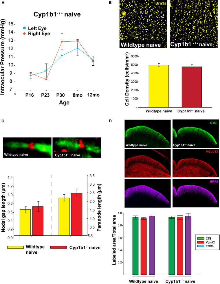
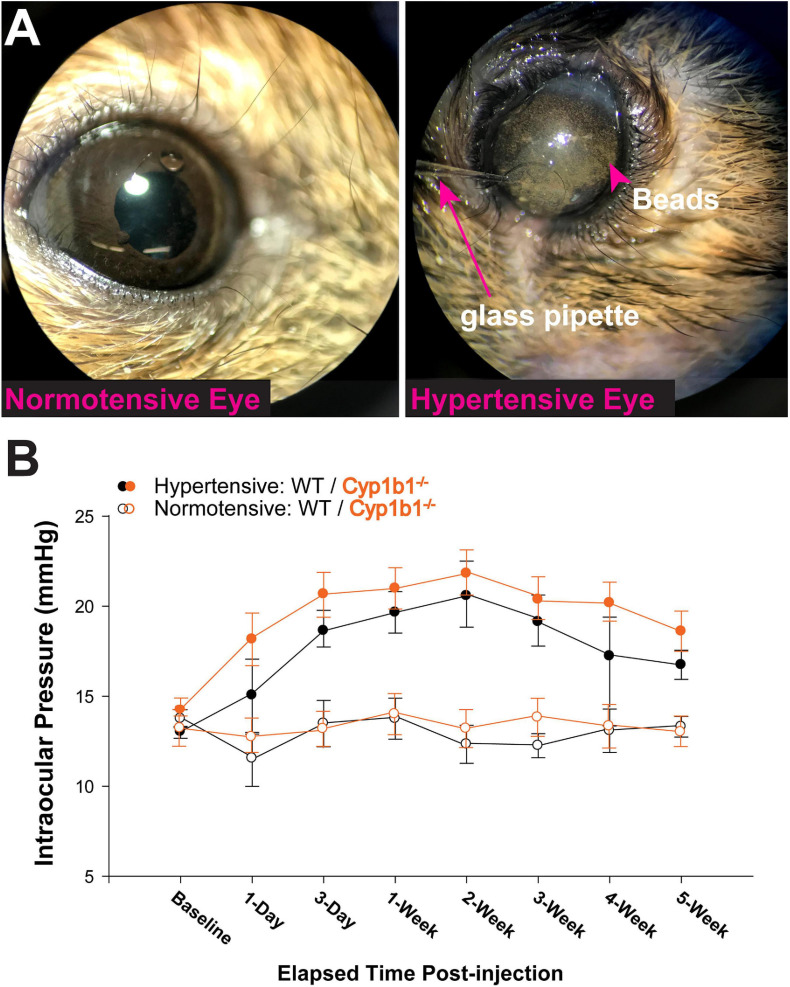
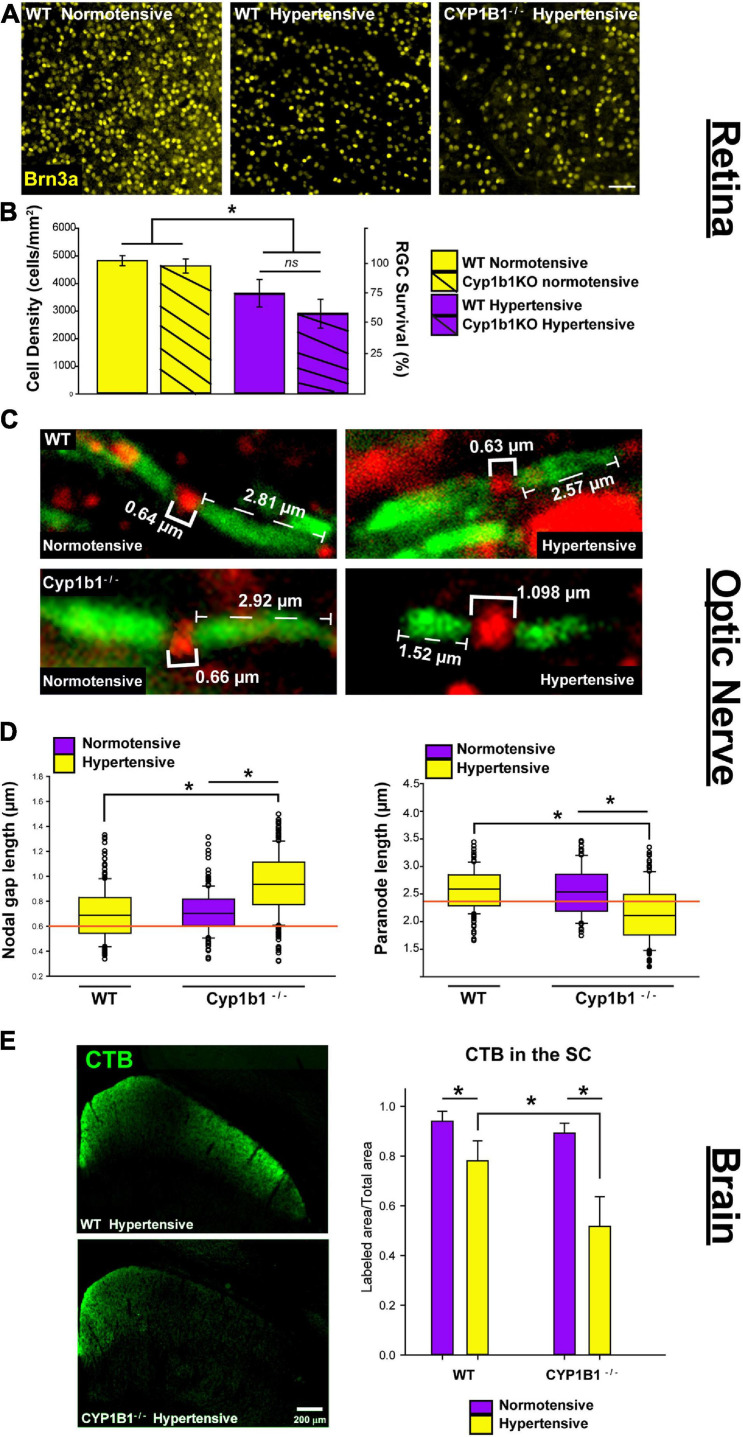
Mutations in the cytochrome P450-1B1 (Cyp1b1) gene is a common genetic predisposition associated with various human glaucomas, most prominently in primary congenital glaucoma (PCG). The role of Cyp1b1 in the eye is largely unknown, however, its absence appears to drive the maldevelopment of anterior eye structures responsible for aqueous fluid drainage in murine models. Nevertheless, vision loss in glaucoma ultimately results from the structural and functional loss of retinal ganglion cells (RGCs). Cyp1b1's influence in the development and support of retinal ganglion cell structure and function under normal conditions or during stress, such as elevated ocular pressure; the most common risk factor in glaucoma, remains grossly unknown. Thus, to determine the role of Cyp1b1 in normal retinal projection development we first assessed the strucutrual integrity of RGCs in the retina, optic nerve, and superior colliculus in un-manipulated (naïve) Cyp1b1-knockout (Cyp1b1-/-) mice. In addition, in a separate cohort of Cyp1b1-/- and wildtype mice, we elevated and maintained intraocular pressure (IOP) at glaucomatous levels for 5-weeks, after which we compared RGC density, node of Ranvier morphology, and axonal transport between the genotypes. Our results demonstrate that naïve Cyp1b1-/- mice develop an anatomically intact retinal projection absent of overt glaucomatous pathology. Following pressure elevation, Cyp1b1-/- accelerated degradation of axonal transport from the retina to the superior colliculus and altered morphology of the nodes of Ranvier and adjacent paranodes in the optic nerves. Together this data suggests the absence Cyp1b1 expression alone is insufficient to drive murine glaucomatous pathology, however, may increase the vulnerability of retinal axons to disease relevant elevations in IOP.

Keywords: axonal transport disruption; glaucoma; microbead occlusion model; nodes of Ranvier; retinal ganglion cell.


Conflict of interest statement

The authors declare that the research was conducted in the absence of any commercial or financial relationships that could be construed as a potential conflict of interest.

Figures

FIGURE 1
FIGURE 1
Cyp1b1–/– develop structurally intact visual projections. (A) Cyp1b1–/– ocular pressure readings across the lifespan (P16 to 12-mo, n = 6) maintained within normal physiological range in both eyes. (B) Retinal ganglion cell density in naïve wildtype vs. Cyp1b1–/– mice. High magnification retinal whole-mount images immunostained for Brn3a, a marker for RGC nuclei. Naïve Cyp1b1–/– (red; n = 12) RGC densities do not differ from naïve wildtype retina (yellow; n = 12). (C) Cyp1b1–/– nodes of Ranvier appear absent of major morphometric changes in the node (Nav1.6, red; n = 200) and paranode (Caspr, green; n = 200). (D) A 50 μm coronal cross section through the superior colliculus of a Cyp1b1–/– mouse that received an intravitreal injection of cholera toxin-B conjugated –alexafluor 488 (CTB488, green) and immunostained for VGlut2 (red, RGC synapses) and estrogen related receptor-β (magenta, RGC axon + axon terminals). Graph depicts superior colliculus label density (labeled area/total area) for each marker. No differences in label density were present between Cyp1b1–/– and wildtype (n = 24).
FIGURE 2
FIGURE 2
Microbead occlusion model. (A) Example image of procedure being performed. Left shows saline injected eye, right shows glass pipette insertion into anterior chamber of subject delivering a solution containing 8 μm magnetic microbeads. (B) Intraocular pressure (IOP) readings from Cyp1b1–/– subjects (orange lines) and wildtype (black lines) following injection of microbeads (hypertensive and solid circles) or saline (normotensive and open circles) over 5 weeks (n = 24).
FIGURE 3
FIGURE 3
Cyp1b1–/– accelerates axonopathy in the retinal projections following ocular pressure elevation. (A) Brn3a immunofluorescence in normotensive and hypertensive flat mount retina from wildtype and Cyp1b1–/– subjects (scale bar = 50 μm). (B) Hypertensive Cyp1b1–/– (solid magenta) and wildtype (dashed magenta) retina demonstrate a significant reduction in Brn3a density compared to normotensive wildtype (solid yellow) and Cyp1b1–/– (dashed yellow) retina. Hypertensive Cyp1b1–/– demonstrate no greater loss in Brn3a density compared to hypertensive wildtype retina. (C) Longitudinal optic nerve sections immunolabeled for Nav1.6 (red) and Caspr (green) to visualize nodes of Ranvier and adjacent paranode in normotensive and hypertensive Cyp1b1–/– and wildtype optic nerves. Brackets indicate node and paranode length in microns. (D) Node lengths increased (left) while paranode lengths decreased (right) in hypertensive (yellow) Cyp1b1–/– optic nerves following 5-week elevation in IOP compared to normotensive (magenta) nerves corresponding to the saline injected eye in the same animal. Nodes and paranode lengths associated with the hypertensive wildtype optic nerves were comparable to wildtype naïve (orange line; indicates average) and normotensive Cyp1b1–/– (magenta) lengths. (E) Epifluorescent images contrasting CTB label in unilateral coronal sections through the superior colliculus of hypertensive Cyp1b1–/– and wildtype subjects. Superior colliculi corresponding to hypertensive eyes (yellow) in both Cyp1b1–/– and wildtype subjects demonstrate significant deficits in axonal transport evidenced by reduced CTB labels compared to colliculi corresponding to the saline injected normotensive eye (magenta). Collicular CTB drop-out was more pronounced in Cyp1b1–/– subjects compared to wildtypes following equivalent period of IOP elevation. Asterisk denotes statistical significance (p < 0.01).

Similar articles

Cited by

References

    1. Akarsu A. N., Turacli M. E., Aktan S. G., Barsoum-Homsy M., Chevrette L., Sayli B. S., et al. (1996). A second locus (GLC3B) for primary congenital glaucoma (Buphthalmos) maps to the 1p36 region. Hum. Mol. Genet. 5 1199–1203. - PubMed
    1. Angelova P. R., Müller W. S. (2009). Arachidonic acid potently inhibits both postsynaptic-type Kv4.2 and presynaptic-type Kv1.4 IA potassium channels. Eur. J. Neurosci. 29 1943–1950. 10.1111/j.1460-9568.2009.06737.x - DOI - PubMed
    1. Aponte E. P., Diehl N., Mohney B. G. (2010). Incidence and clinical characteristics of childhood glaucoma: a population-based study. Arch. Ophthalmol. 128 478–482. 10.1001/archophthalmol.2010.41.Incidence - DOI - PMC - PubMed
    1. Baltan S., Inman D. M., Danilov C. A., Morrison R. S., Calkins D. J., Horner P. J. (2010). Metabolic vulnerability disposes retinal ganglion cell axons to dysfunction in a model of glaucomatous degeneration. J. Neurosci. 30 5644–5652. 10.1523/JNEUROSCI.5956-09.2010 - DOI - PMC - PubMed
    1. Bansal S., Leu A. N., Gonzalez F. J., Guengerich F. P., Chowdhury A. R., Anandatheerthavarada H. K., et al. (2014). Mitochondrial targeting of cytochrome P450 (CYP) 1B1 and its role in polycyclic aromatic hydrocarbon-induced mitochondrial dysfunction. J. Biol. Chem. 289 9936–9951. 10.1074/jbc.M113.525659 - DOI - PMC - PubMed

LinkOut - more resources