Myocardial Infarction: The Protective Role of MiRNAs in Myocardium Pathology
- PMID: 33748196
- PMCID: PMC7973051
- DOI: 10.3389/fcvm.2021.631817
Myocardial Infarction: The Protective Role of MiRNAs in Myocardium Pathology
Abstract
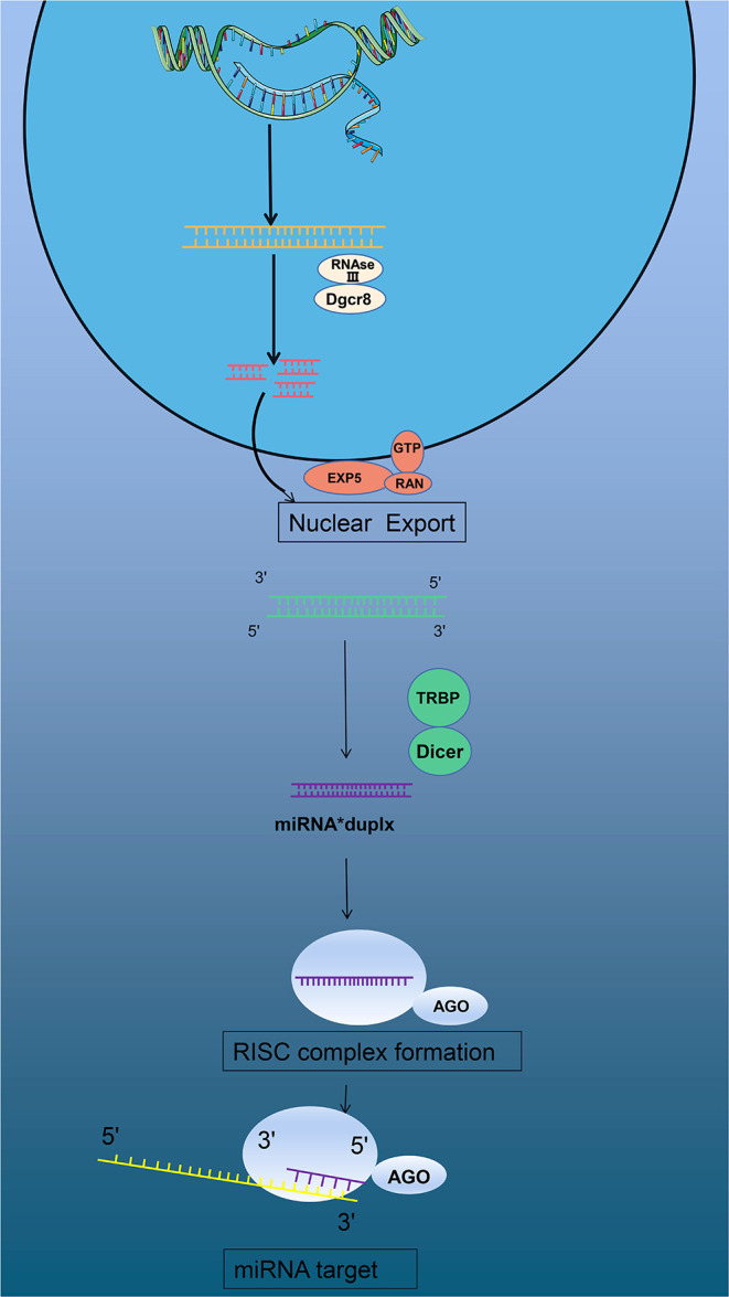
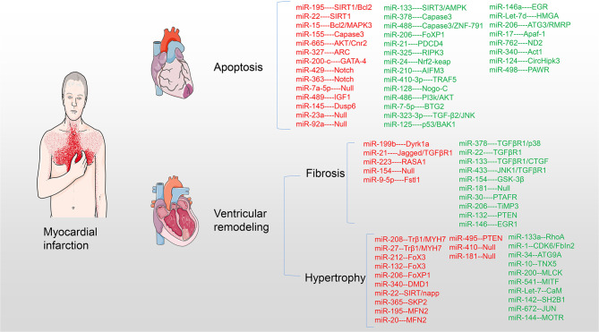
Cardiovascular diseases have been regarded as the leading cause of death around the world, with myocardial infarction (MI) being the most severe form. MI leads to myocardial apoptosis, cardiomyocyte fibrosis, and cardiomyocyte hypertrophy, ultimately leading to heart failure, and death. Micro RNAs (miRNAs) participate in the genesis and progression of myocardial pathology after MI by playing an important regulatory role. This review aims to summarize all available knowledge on the role of miRNAs in the myocardial pathological process after MI to uncover potential major target pathways. In addition, the main therapeutic methods and their latest progress are also reviewed. miRNAs can regulate the main signaling pathways as well as pathological processes. Thus, they have the potential to induce therapeutic effects. Hence, the combination of miRNAs with recently developed exosome nanocomplexes may represent the future direction of therapeutics.
Keywords: MI; apoptosis; fibrosis; hypertrophy; miRNA.
Copyright © 2021 Wang and Zheng.
Conflict of interest statement
The authors declare that the research was conducted in the absence of any commercial or financial relationships that could be construed as a potential conflict of interest.
Figures


Similar articles
-
The emerging role of miRNAs in myocardial infarction: From molecular signatures to therapeutic targets.Pathol Res Pract. 2024 Jan;253:155087. doi: 10.1016/j.prp.2023.155087. Epub 2024 Jan 3. Pathol Res Pract. 2024. PMID: 38183820 Review.
-
miRNome in myocardial infarction: Future directions and perspective.World J Cardiol. 2014 Sep 26;6(9):939-58. doi: 10.4330/wjc.v6.i9.939. World J Cardiol. 2014. PMID: 25276296 Free PMC article. Review.
-
The Regulation Mechanisms and Clinical Application of MicroRNAs in Myocardial Infarction: A Review of the Recent 5 Years.Front Cardiovasc Med. 2022 Jan 17;8:809580. doi: 10.3389/fcvm.2021.809580. eCollection 2021. Front Cardiovasc Med. 2022. PMID: 35111829 Free PMC article. Review.
-
miRNAs Epigenetic Tuning of Wall Remodeling in the Early Phase after Myocardial Infarction: A Novel Epidrug Approach.Int J Mol Sci. 2023 Aug 26;24(17):13268. doi: 10.3390/ijms241713268. Int J Mol Sci. 2023. PMID: 37686073 Free PMC article. Review.
-
MicroRNAs in the development of left ventricular remodeling and postmyocardial infarction heart failure.Pol Arch Intern Med. 2020 Jan 31;130(1):59-65. doi: 10.20452/pamw.15137. Epub 2020 Jan 14. Pol Arch Intern Med. 2020. PMID: 31933487 Review.
Cited by
-
Deficiency of miR-409-3p improves myocardial neovascularization and function through modulation of DNAJB9/p38 MAPK signaling.Mol Ther Nucleic Acids. 2023 May 20;32:995-1009. doi: 10.1016/j.omtn.2023.05.021. eCollection 2023 Jun 13. Mol Ther Nucleic Acids. 2023. PMID: 37332476 Free PMC article.
-
MicroRNA-409-3p/BTG2 signaling axis improves impaired angiogenesis and wound healing in obese mice.FASEB J. 2024 Feb 15;38(3):e23459. doi: 10.1096/fj.202302124RR. FASEB J. 2024. PMID: 38329343 Free PMC article.
-
Plasma Exosomes at the Late Phase of Remote Ischemic Pre-conditioning Attenuate Myocardial Ischemia-Reperfusion Injury Through Transferring miR-126a-3p.Front Cardiovasc Med. 2021 Nov 30;8:736226. doi: 10.3389/fcvm.2021.736226. eCollection 2021. Front Cardiovasc Med. 2021. PMID: 34917657 Free PMC article.
-
Advances in the study of exosomes derived from mesenchymal stem cells and cardiac cells for the treatment of myocardial infarction.Cell Commun Signal. 2023 Aug 14;21(1):202. doi: 10.1186/s12964-023-01227-9. Cell Commun Signal. 2023. PMID: 37580705 Free PMC article. Review.
-
An exploration into the diagnostic capabilities of microRNAs for myocardial infarction using machine learning.Biol Direct. 2024 Dec 10;19(1):127. doi: 10.1186/s13062-024-00543-5. Biol Direct. 2024. PMID: 39658789 Free PMC article.
References
Publication types
LinkOut - more resources
Full Text Sources
Other Literature Sources