. 2021 Mar 4:8:639752.
doi: 10.3389/fvets.2021.639752. eCollection 2021.

Changes in Oviductal Cells and Small Extracellular Vesicles miRNAs in Pregnant Cows

Affiliations

Changes in Oviductal Cells and Small Extracellular Vesicles miRNAs in Pregnant Cows

Rosane Mazzarella et al. Front Vet Sci. .

Abstract

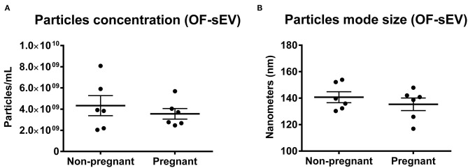
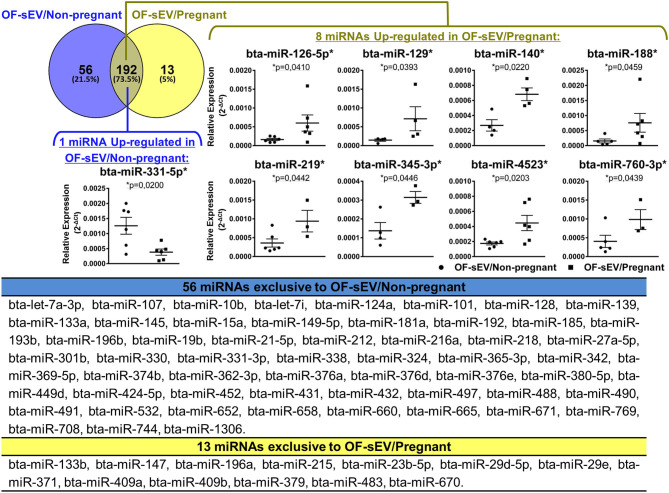
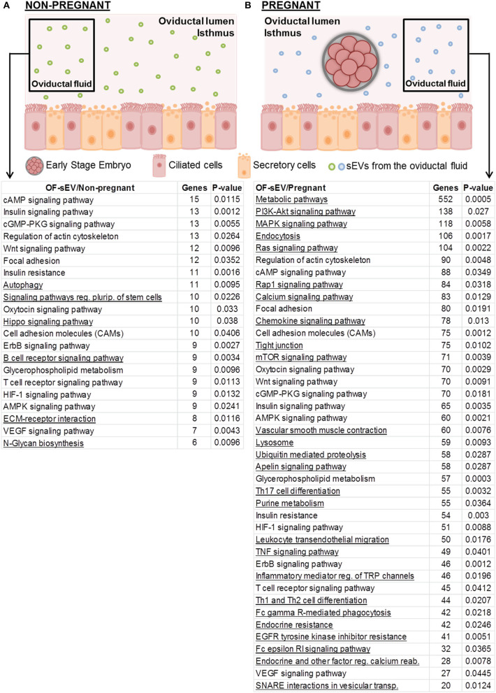
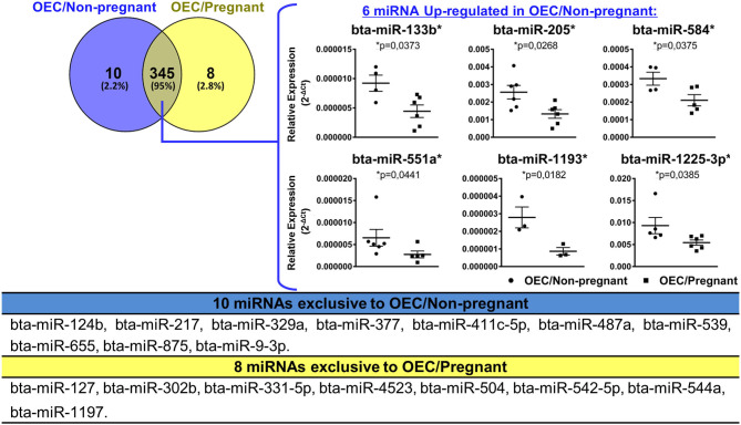
Early embryonic development occurs in the oviduct, where an ideal microenvironment is provided by the epithelial cells and by the oviductal fluid produced by these cells. The oviductal fluid contains small extracellular vesicles (sEVs), which through their contents, including microRNAs (miRNAs), can ensure proper cell communication between the mother and the embryo. However, little is known about the modulation of miRNAs within oviductal epithelial cells (OECs) and sEVs from the oviductal fluid in pregnant cows. In this study, we evaluate the miRNAs profile in sEVs from the oviductal flushing (OF-sEVs) and OECs from pregnant cows compared to non-pregnant, at 120 h after ovulation induction. In OF-sEVs, eight miRNAs (bta-miR-126-5p, bta-miR-129, bta-miR-140, bta-miR-188, bta-miR-219, bta-miR-345-3p, bta-miR-4523, and bta-miR-760-3p) were up-regulated in pregnant and one miRNA (bta-miR-331-5p) was up-regulated in non-pregnant cows. In OECs, six miRNAs (bta-miR-133b, bta-miR-205, bta-miR-584, bta-miR-551a, bta-miR-1193, and bta-miR-1225-3p) were up-regulated in non-pregnant and none was up-regulated in pregnant cows. Our results suggest that embryonic maternal communication mediated by sEVs initiates in the oviduct, and the passage of gametes and the embryo presence modulate miRNAs contents of sEVs and OECs. Furthermore, we demonstrated the transcriptional levels modulation of selected genes in OECs in pregnant cows. Therefore, the embryonic-maternal crosstalk potentially begins during early embryonic development in the oviduct through the modulation of miRNAs in OECs and sEVs in pregnant cows.

Keywords: bovine; embryo maternal-communication; miRNAs; oviductal cells; oviductal fluid; reproduction; small extracellular vesicles.


Conflict of interest statement

The authors declare that the research was conducted in the absence of any commercial or financial relationships that could be construed as a potential conflict of interest.

Figures

Figure 1
Figure 1
Characterization of OF-sEVs by nanoparticle analysis. Nanoparticle tracking analysis showing no difference in particles concentration (A) and mode size (B) between the groups OF-sEV/Non-pregnant (n = 6) and OF-sEV/Pregnant (n = 6). Error bars represents standard errors of the means.
Figure 2
Figure 2
miRNAs profile of OF-sEVs isolated from non-pregnant and pregnant cows. Venn diagram representing the 261 miRNAs detected: 192 in common between the two groups, 56 exclusive to OF-sEV/Non-pregnant and 13 exclusive to OF-sEV/Pregnant. Among the 192 miRNAs detected in both groups, eight miRNAs (bta-miR-126-5p, bta-miR-129, bta-miR-140, bta-miR-188, bta-miR-219, bta-miR-345-3p, bta-miR-4523, and bta-miR-760-3p) were up-regulated in OF-sEV/Pregnant and one miRNA (bta-miR-331-5p) was up-regulated in OF-sEV/Non-pregnant. The asterisk (*) indicates miRNAs with relative expression significantly different (p < 0.05) within the groups. Samples size were n = 6 for both groups. Error bars represent SEM.
Figure 3
Figure 3
Selected biological pathways predicted to be regulated by miRNAs with increased expression in OF-sEVs. (A) Pathways predicted as regulated by the miRNA (bta-miR-331-5p) up-regulated in OF-sEV/Non-pregnant. (B) Pathways predicted as regulated by the eight miRNAs (bta-miR-126-5p, bta-miR-129, bta-miR-140, bta-miR-188, bta-miR-219, bta-miR-345-3p, bta-miR-4523, and bta-miR-760-3p) up-regulated in OF-sEV/Pregnant. Pathways were selected based on the potential relevance for oviductal functions and p < 0.05. Underlined pathways are modulated only in the group that they are present.
Figure 4
Figure 4
miRNAs profile of OECs from non-pregnant and pregnant cows. Venn diagram representing the detection of 363 miRNAs: 345 common between the two groups, 10 exclusive to OEC/Non-pregnant and eight exclusive to OEC/Pregnant. Among the 345 miRNAs detected in both groups, six miRNAs (bta-miR-133b, bta-miR-205, bta-miR-584, bta-miR-551a, bta-miR-1193, and bta-miR-1225-3p) were up-regulated in OEC/Non-pregnant. The asterisk (*) indicates miRNAs with relative expression significantly different (p < 0.05) within the groups. Samples size were n = 6 for both groups. Error bars represent SEM.
Figure 5
Figure 5
Biological pathways predicted using up-regulated miRNAs in OECs. Pathways predicted as regulated by six miRNAs (bta-miR-133b, bta-miR-205, bta-miR-584, bta-miR-551a, bta-miR-1193, and bta-miR-1225-3p) up-regulated in OECs from non-pregnant compared with OECs from pregnant cows. The left Y-axis values represent the number of genes predicted as modulated by the miRNAs for the respective pathways. The right Y-axis represents the enrichment score (–log10 of the p-value) for each pathway.
Figure 6
Figure 6
Relative mRNA expression of selected genes in OECs from non-pregnant compared with pregnant cows. Relative expression of up and downregulated mRNAs are shown as mean of 2−ΔCt ± SEM. One asterisk (*) indicates genes with p < 0.05 and two asterisks (**) indicates genes with p < 0.1 within the groups. Samples size were n = 6 for both groups.

Similar articles

Cited by

References

    1. Besenfelder U, Havlicek V, Brem G. Role of the oviduct in early embryo development. Reprod Domest Anim. (2012) 47:156–63. 10.1111/j.1439-0531.2012.02070.x - DOI - PubMed
    1. Avilés M, Coy P, Rizos D. The oviduct: a key organ for the success of early reproductive events. Anim Front. (2015) 5:25–31. 10.2527/af.2015-0005 - DOI
    1. Kölle S, Dubielzig S, Reese S, Wehrend A, König P, Kummer W. Ciliary transport, gamete interaction, and effects of the early embryo in the oviduct: ex vivo analyses using a new digital videomicroscopic system in the cow. Biol Reprod. (2009) 81:267–74. 10.1095/biolreprod.108.073874 - DOI - PubMed
    1. Leese HJ, Hugentobler SA, Gray SM, Morris DG, Sturmey RG, Whitear SL, et al. . Female reproductive tract fluids: composition, mechanism of formation and potential role in the developmental origins of health and disease. Reprod Fertil Dev. (2008) 20:1–8. 10.1071/RD07153 - DOI - PubMed
    1. Avilés M, Gutiérrez-Adán A, Coy P. Oviductal secretions: will they be key factors for the future ARTs? Mol Hum Reprod. (2010) 16:896–906. 10.1093/molehr/gaq056 - DOI - PubMed