

Focal Adhesion Kinase Inhibitor Inhibits the Oxidative Damage Induced by Central Venous Catheter via Abolishing Focal Adhesion Kinase-Protein Kinase B Pathway Activation
- PMID: 33748278
- PMCID: PMC7943296
- DOI: 10.1155/2021/6685493
Focal Adhesion Kinase Inhibitor Inhibits the Oxidative Damage Induced by Central Venous Catheter via Abolishing Focal Adhesion Kinase-Protein Kinase B Pathway Activation
Abstract
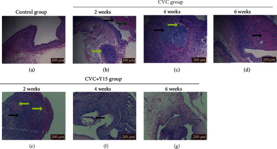
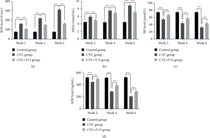
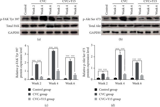
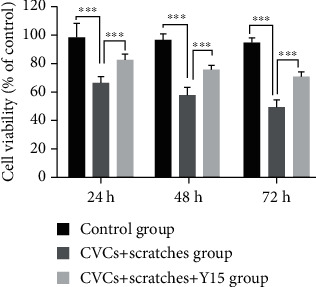
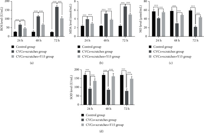
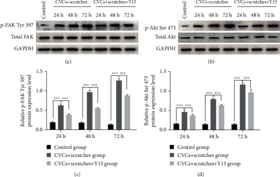
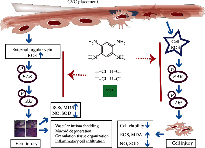
The vascular injury induced by central venous catheter (CVC) indwelling is the basis for the occurrence and development of CVC-related complications, such as phlebitis, venous thrombosis, and catheter-related infections. Focal adhesion kinase (FAK) and FAK-protein kinase B (AKT) signaling pathway are of great significance in tissue repair after trauma. Here, we investigated the role and mechanism of the FAK inhibitor (1,2,4,5-phenyltetramine tetrahydrochloride (Y15)) in oxidative damage caused by CVC. EA.hy926 cells were divided into the control group (normal control), CVCs+scratches group (the intercepted CVC segments coculturing with scratched EA.hy926 cells), and CVCs+scratches+Y15 group (Y15 was added to the cell culture supernatant with CVCs + scratches at a final concentration of 50 μmol·L-1). New Zealand rabbits were randomly divided into the control group (normal control), CVC group (CVC was inserted through the rabbit's right jugular vein to the junction of the right atrium and superior vena cava), and CVC+Y15 group (CVC was immersed in a 50 μmol·L-1 Y15 solutions before insertion). The levels of markers and proteins related to oxidative damage in cells, cell culture supernatant, serum, and external jugular vein were measured by commercial kits and western blot, respectively. We found that Y15 treatment significantly decreased ROS and MDA levels and increased cell viability, NO, and SOD levels in a time-dependent manner in rabbit serum and cell culture supernatant. In addition, Y15 effectively reduced the CVC-induced pathological changes of damaged vascular tissues. Y15 also downregulated the levels of p-FAK Tyr 397 and p-Akt Ser 473 in damaged external jugular vein and EA.hy926 cells. These findings suggest that Y15 alleviated CVC-induced oxidative damage to blood vessels by suppressing focal FAK-Akt pathway activation.
Copyright © 2021 Yanru Wang et al.
Conflict of interest statement
The authors declare that there is no conflict of interest regarding the publication of this paper.
Figures
Similar articles
-
FAK inhibition with small molecule inhibitor Y15 decreases viability, clonogenicity, and cell attachment in thyroid cancer cell lines and synergizes with targeted therapeutics.Oncotarget. 2014 Sep 15;5(17):7945-59. doi: 10.18632/oncotarget.2381. Oncotarget. 2014. PMID: 25277206 Free PMC article.
-
In vivo toxicity, metabolism and pharmacokinetic properties of FAK inhibitor 14 or Y15 (1, 2, 4, 5-benzenetetramine tetrahydrochloride).Arch Toxicol. 2015 Jul;89(7):1095-101. doi: 10.1007/s00204-014-1290-y. Epub 2014 Jun 12. Arch Toxicol. 2015. PMID: 24915938
-
Inhibition of cell migration by focal adhesion kinase: Time-dependent difference in integrin-induced signaling between endothelial and hepatoblastoma cells.Int J Mol Med. 2018 May;41(5):2573-2588. doi: 10.3892/ijmm.2018.3512. Epub 2018 Feb 23. Int J Mol Med. 2018. PMID: 29484384 Free PMC article.
-
Fourteen years of hemodialysis with a central venous catheter: mechanical long-term complications.J Vasc Access. 2006 Apr-Jun;7(2):60-5. doi: 10.1177/112972980600700204. J Vasc Access. 2006. PMID: 16868898 Review.
-
Repair of Central Venous Catheters in Home Parenteral Nutrition Patients.Nutr Clin Pract. 2019 Apr;34(2):210-215. doi: 10.1002/ncp.10262. Epub 2019 Feb 7. Nutr Clin Pract. 2019. PMID: 30729597 Review.
Cited by
-
Chicken Primordial Germ Cells Do Not Proliferate in Insulin-Lacking Media.Int J Mol Sci. 2025 Mar 28;26(7):3122. doi: 10.3390/ijms26073122. Int J Mol Sci. 2025. PMID: 40243906 Free PMC article.
-
Progress in Research on the Mechanisms and Interventions of Phlebitis from the Perspective of Vascular Endothelial Cell and Signaling Pathway.J Inflamm Res. 2023 Dec 29;16:6469-6481. doi: 10.2147/JIR.S450149. eCollection 2023. J Inflamm Res. 2023. PMID: 38170089 Free PMC article. Review.
References
MeSH terms
Substances
LinkOut - more resources
Full Text Sources
Other Literature Sources
Miscellaneous
