

Influence of Piper betle L. extract on umbilical cord cells in vitro and potential treating cutaneous wound
- PMID: 33748448
- PMCID: PMC7969898
- DOI: 10.1016/j.heliyon.2021.e06248
Influence of Piper betle L. extract on umbilical cord cells in vitro and potential treating cutaneous wound
Abstract
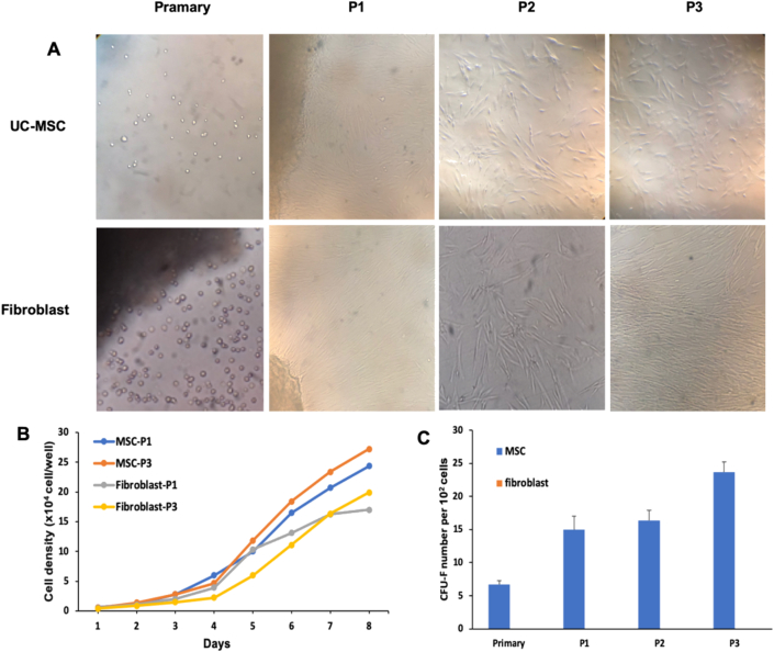
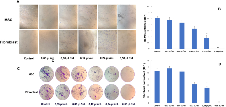
This research aimed to test the effects of Piper betle L. from Vietnam on fibroblasts and UC-derived mesenchymal stem cells (UC-MSCs) from human umbilical cord (UC) on the scratch assay. We tested the extract at different concentrations and then assessed the level of expression of the factors involved in the inflammatory process on fibroblasts including IL-33, VCAM, CD248 by assay real time qPCR. At the concentrations of 0.025 μL/mL and 0.03 μL/mL, the extracts positively affected fibroblast proliferation and UC-MSCs. By contrast, the concentration of 0.058 μL/mL, the extract was toxic to UC-MSCs and fibroblast cell lines, the cells were no longer able to survive. qPCR results show that Piper betle L. extract has the ability to reduce the expression levels of IL-33 (50.8%), VCAM (32.1%), CD248 (46.13%) which trigger inflammatory processes, thereby reducing cellular stress and promoting the process of healing scratches. Our study revealed the proliferation capabilities of Piper betle L. extract from Vietnam In vitro. Hence, Piper betle L. could be recommended as a potential source of wound healing agents.
Keywords: Fibroblast cell; Mesenchymal stem cells; Piper betle L.; The extract.
© 2021 The Authors.
Conflict of interest statement
The authors declare no conflict of interest.
Figures


References
-
- Li J., Chen J., Kirsner R. Pathophysiology of acute wound healing. Clin. Dermatol. 2007 Jan 1;25(1):9–18. - PubMed
-
- Bainbridge P. Wound healing and the role of fibroblasts. J. Wound Care. 2013;22(8):407–412. - PubMed
-
- Buckley C.D., Gilroy D.W., Serhan C.N., Stockinger B., Tak P.P. The resolution of inflammation. Nat. Rev. Immunol. 2013 Jan;13(1):59–66. - PubMed
LinkOut - more resources
Full Text Sources
Other Literature Sources
Miscellaneous
