. 2021 Mar 10;2(1):100370.
doi: 10.1016/j.xpro.2021.100370. eCollection 2021 Mar 19.

Co-fractionation/mass spectrometry to identify protein complexes

Affiliations

Co-fractionation/mass spectrometry to identify protein complexes

Claire D McWhite et al. STAR Protoc. .

Abstract

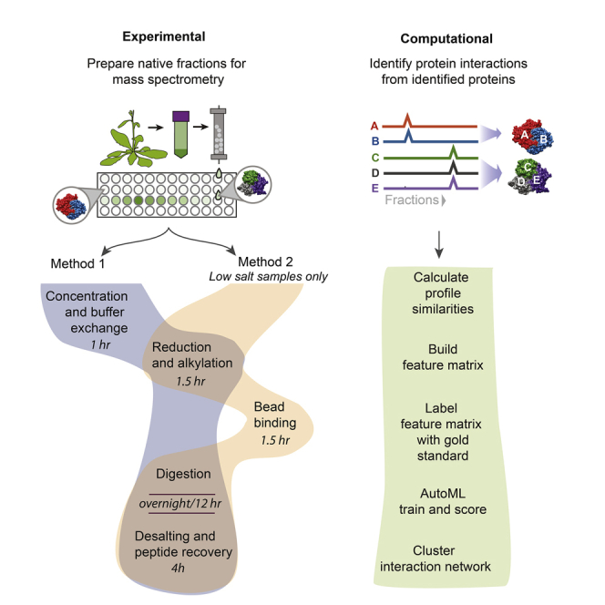
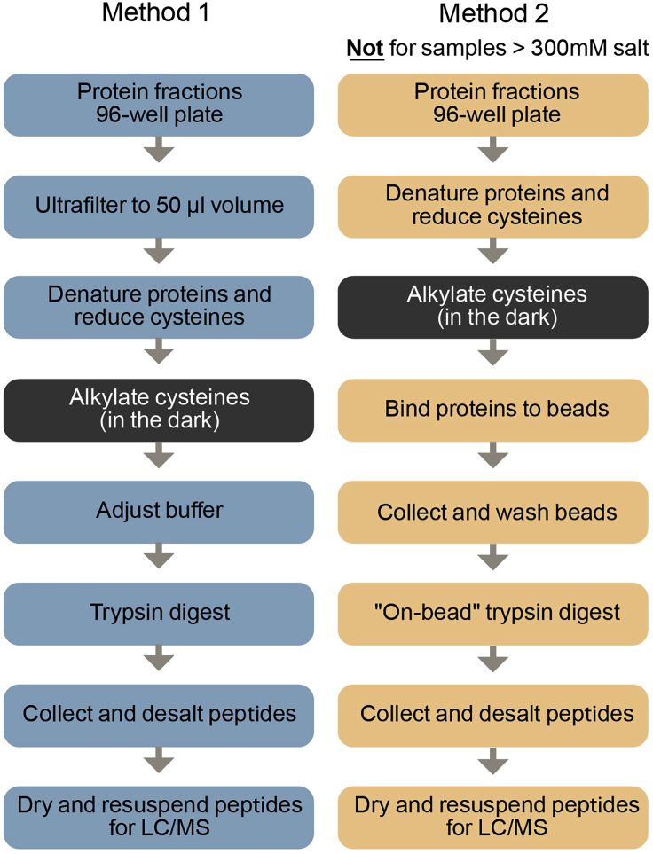
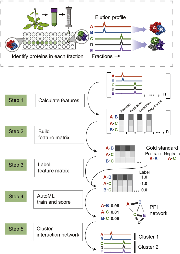
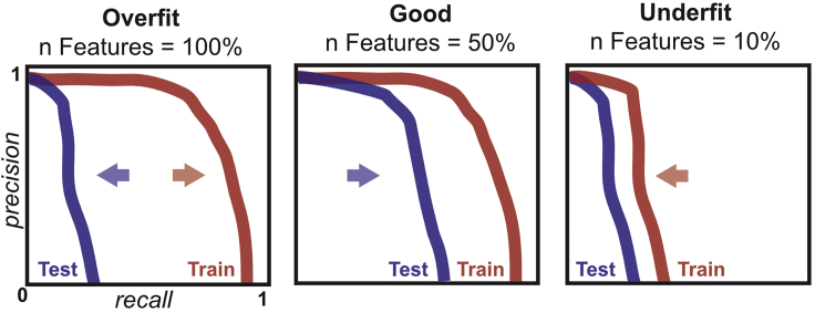
Co-fractionation/mass spectrometry (CF/MS) is a flexible and powerful method to detect physical associations of proteins. CF/MS can be applied to any tissue or organism without the need for protein-specific antibodies or epitope tags. Here, we outline two alternate protocols for MS preparation of samples (containing low or high salt) and a computational pipeline (cfmsflow) that together allow the successful application of this approach. These protocols are based on CF/MS of over 16 diverse organisms including plants and animals. For complete details on the use and execution of this protocol, please refer to McWhite et al. (2020).

Keywords: Bioinformatics; Proteomics.


Conflict of interest statement

The authors have no competing interests.

Figures

None
Graphical abstract
Figure 1
Figure 1
Workflow of the two alternative methods for preparation of LC/MS-ready samples from chromatographic fractions Method 1 can be used in any salt condition but Method 2 will only work if salt is below 300 mM.
Figure 2
Figure 2
Modification of a 96-well fraction collection plate (shown here, Corning Axygen polypropylene #P-DW-20-C) for use with the 96-well magnetic plate separator (Thermo Fisher Scientific #A14179) Four support ribs on the underside of the deep well plate (indicated by black arrows in lower left) interfere with prongs on the blue magnetic plate preventing close contact of the well bottoms with the magnet. The 4 ribs are cut and folded as follows to accommodate the prongs. For each of the 4 interfering support ribs use a straight-edge razor to make 2 vertical cuts of the approximate depth shown. Use metal forceps or other tool to bend the resultant plastic tabs over. After modification of the 4 ribs the deepwell plate should sit flush on the magnetic plate as shown in the upper right panel.
Figure 3
Figure 3
Overview of computational pipeline to detect protein-protein interactions and protein complexes In step 1, a set of similarity scores between all proteins are calculated for each fractionation experiment. In step 2, these similarity scores are combined into one large table. In step 3, pairs of proteins that are known from prior literature to interact are labeled with a 1 (positive training label), and a set of random pairs of proteins are labeled with a −1 (negative training label). In step 4, a model is trained to distinguish these positive and negatively labeled pairs of proteins, giving a score to each pair, where a higher score indicates higher probability of interaction. In step 5, this interaction network is clustered to protein complexes.
Figure 4
Figure 4
Using precision-recall curves to evaluate overfitting Precision-recall curves illustrating overfit, better fit, and underfit models. A substantial difference between Test and Train precision-recall curves suggests overfitting, and that the max number of features should likely be lowered to improve model performance. When a model is overfit, as features are removed, the Test precision-recall curve will shift right, while the training curve is minimally affected. Once too many features have been removed, performance in both test and training will decline.

References

    1. Di Tommaso P., Chatzou M., Floden E.W., Barja P.P., Palumbo E., Notredame C. Nextflow enables reproducible computational workflows. Nat. Biotechnol. 2017;35:316–319. - PubMed
    1. Drew K., Lee C., Cox R.M., Dang V., Devitt C.C., Papoulas O., Huizar R.L., Marcotte E.M., Wallingford J.B. A systematic, label-free method for identifying RNA-associated proteins in vivo provides insights into vertebrate ciliary beating machinery. Dev. Biol. 2020;467:108–117. - PMC - PubMed
    1. Drew K., Lee C., Huizar R.L., Tu F., Borgeson B., McWhite C.D., Ma Y., Wallingford J.B., Marcotte E.M. Integration of over 9,000 mass spectrometry experiments builds a global map of human protein complexes. Mol. Syst. Biol. 2017;13(6):932. doi: 10.15252/msb.20167490. - DOI - PMC - PubMed
    1. Liebeskind B.J., Young R.L., Halling D.B., Aldrich R.W., Marcotte E.M. Mapping functional protein neighborhoods in the mouse brain. bioRxiv. 2020 doi: 10.1101/2020.01.26.920447. - DOI
    1. Mallam A.L., Sae-Lee W., Schaub J.M., Tu F., Battenhouse A., Jang Y.J., Kim J., Finkelstein I.J., Marcotte E.M., Drew K. Systematic discovery of endogenous human ribonucleoprotein complexes. Cell Rep. 2019;29:1351–1368.e5. - PMC - PubMed

Publication types

LinkOut - more resources