. 2021 Jan 5;2(1):1-13.
doi: 10.1089/neur.2020.0020. eCollection 2021.

Characterization of the Jet-Flow Overpressure Model of Traumatic Brain Injury in Mice

Affiliations

Characterization of the Jet-Flow Overpressure Model of Traumatic Brain Injury in Mice

Min-Kyoo Shin et al. Neurotrauma Rep. .

Abstract

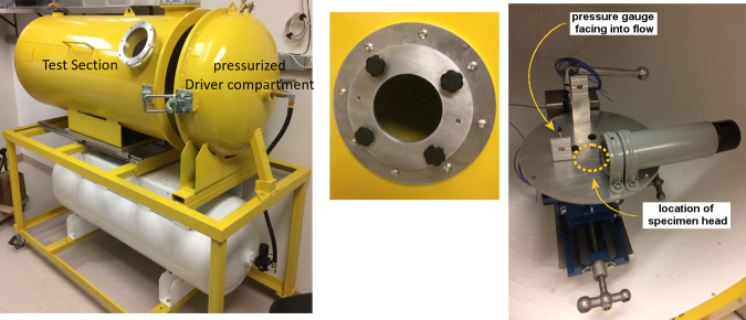
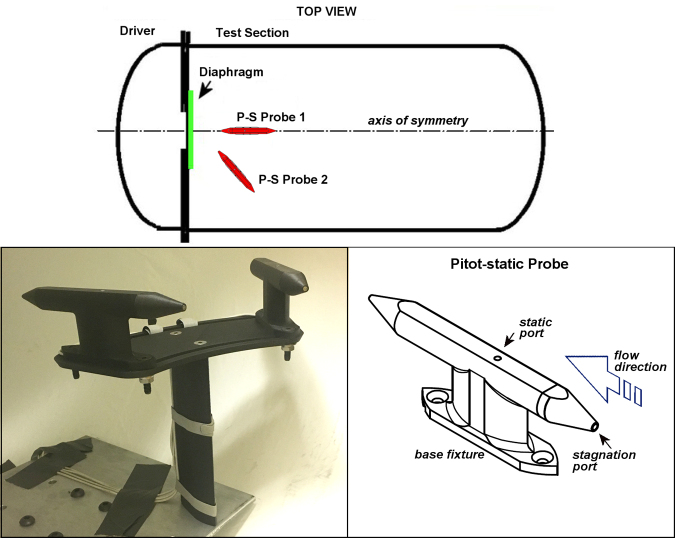
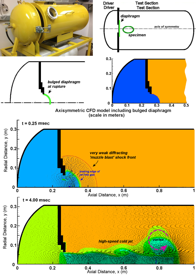
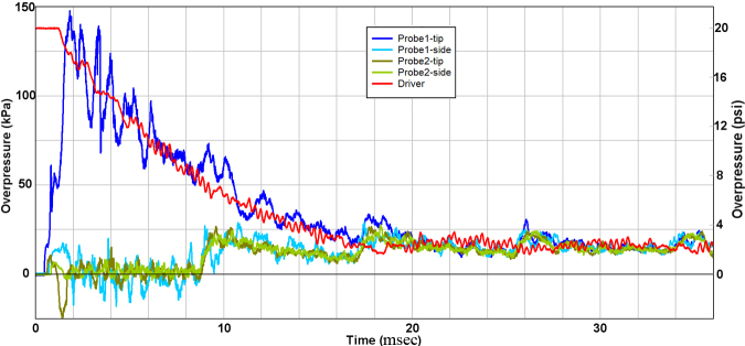
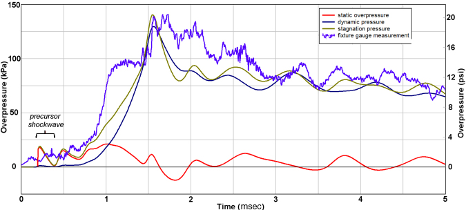
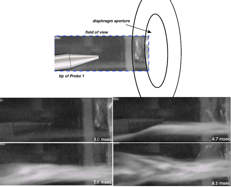
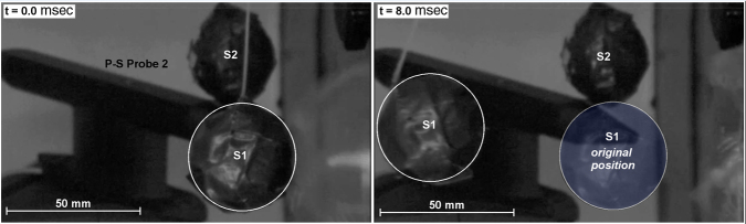
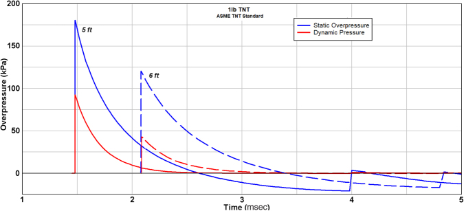
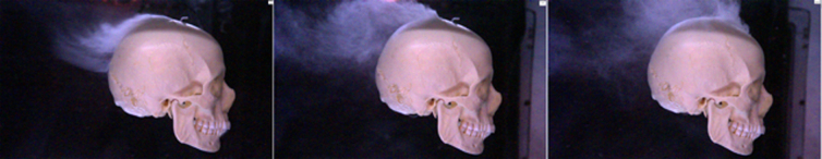
The jet-flow overpressure chamber (OPC) has been previously reported as a model of blast-mediated traumatic brain injury (bTBI). However, rigorous characterization of the features of this injury apparatus shows that it fails to recapitulate exposure to an isolated blast wave. Through combined experimental and computational modeling analysis of gas-dynamic flow conditions, we show here that the jet-flow OPC produces a collimated high-speed jet flow with extreme dynamic pressure that delivers a severe compressive impulse. Variable rupture dynamics of the diaphragm through which the jet flow originates also generate a weak and infrequent shock front. In addition, there is a component of acceleration-deceleration injury to the head as it is agitated in the headrest. Although not a faithful model of free-field blast exposure, the jet-flow OPC produces a complex multi-modal model of TBI that can be useful in laboratory investigation of putative TBI therapies and fundamental neurophysiological processes after brain injury.

Keywords: blast; jet flow; multi-modal traumatic brain injury; overpressure chamber.


Conflict of interest statement

No competing financial interests exist.

Figures

FIG. 1.
FIG. 1.
This is a photograph of the principle components of the jet-flow overpressure chamber. On the left is shown the close-ended Test Section into which the animal is placed, and the Pressurize Drive Compartment into which compressed air is delivered in order to burst the membrane. In the middle is shown the diaphragm where the Mylar membrane is placed, between the two chambers. On the right is shown the holder into which the animal is placed.
FIG. 2.
FIG. 2.
Configuration of the Pitot-static probes installed for analyses of free-field flow conditions generated by the jet-flow overpressure chamber.
FIG. 3.
FIG. 3.
Illustration of the computational fluid dynamics model of the jet-flow overpressure chamber. Animation of the jet-flow development is presented in Supplemental Figure S1.
FIG. 4.
FIG. 4.
Representative experimental records from current work.
FIG. 5.
FIG. 5.
Comparison of experimental records with computational fluid dynamic modeling for the early flow development and peak levels.
FIG. 6.
FIG. 6.
Frame sequence from high-speed imaging of jet-flow developed at the nominal specimen location. For purposes of referencing time relative to the bursting of the diaphragm, 0.11 ms would be added to each time shown. High-speed video is presented in Supplemental Figure S2.
FIG. 7.
FIG. 7.
Frame sequence from high-speed imaging of the free-flight motion of a ballasted sphere subjected to the jet-flow impingement. The test sphere attained a velocity of about 15 m/s within 5 ms or an acceleration about 270g.
FIG. 8.
FIG. 8.
Description of blast-wave profiles for static and dynamic pressure (adapted with permission from Glasstone and Dolan).
FIG. 9.
FIG. 9.
Description of blast-wave profiles for static and dynamic pressure for the case of 1 lb (0.454 kg) TNT at distances of 5 and 6 ft standoff (1.52, 1.83 m). (reprinted with permission from American National Standards Institute).
FIG. 10.
FIG. 10.
Frame sequence from high-speed imaging of blast-wave exposure of ‘free-flight’ human-skull replica showing flow patterns; negligible motion was imparted for a blast of 125 kPa and 6 ms duration. (reprinted with permission from Ritzel and colleagues).

Similar articles

Cited by

  • Reducing acetylated tau is neuroprotective in brain injury.
    Shin MK, Vázquez-Rosa E, Koh Y, Dhar M, Chaubey K, Cintrón-Pérez CJ, Barker S, Miller E, Franke K, Noterman MF, Seth D, Allen RS, Motz CT, Rao SR, Skelton LA, Pardue MT, Fliesler SJ, Wang C, Tracy TE, Gan L, Liebl DJ, Savarraj JPJ, Torres GL, Ahnstedt H, McCullough LD, Kitagawa RS, Choi HA, Zhang P, Hou Y, Chiang CW, Li L, Ortiz F, Kilgore JA, Williams NS, Whitehair VC, Gefen T, Flanagan ME, Stamler JS, Jain MK, Kraus A, Cheng F, Reynolds JD, Pieper AA. Shin MK, et al. Cell. 2021 May 13;184(10):2715-2732.e23. doi: 10.1016/j.cell.2021.03.032. Epub 2021 Apr 13. Cell. 2021. PMID: 33852912 Free PMC article.
  • From imaging to intervention: emerging potential of PET biomarkers to shape therapeutic strategies for TBI-induced neurodegeneration.
    Giarratana AO. Giarratana AO. Front Neurol. 2025 Aug 5;16:1637243. doi: 10.3389/fneur.2025.1637243. eCollection 2025. Front Neurol. 2025. PMID: 40837059 Free PMC article. Review.
  • Acutely blocking excessive mitochondrial fission prevents chronic neurodegeneration after traumatic brain injury.
    Sridharan PS, Koh Y, Miller E, Hu D, Chakraborty S, Tripathi SJ, Kee TR, Chaubey K, Vázquez-Rosa E, Barker S, Liu H, León-Alvarado RA, Franke K, Cintrón-Pérez CJ, Dhar M, Shin MK, Flanagan ME, Castellani RJ, Gefen T, Bykova M, Dou L, Cheng F, Wilson BM, Fujioka H, Kang DE, Woo JA, Paul BD, Qi X, Pieper AA. Sridharan PS, et al. Cell Rep Med. 2024 Sep 17;5(9):101715. doi: 10.1016/j.xcrm.2024.101715. Epub 2024 Sep 5. Cell Rep Med. 2024. PMID: 39241772 Free PMC article.
  • Longitudinal in vivo monitoring of axonal degeneration after brain injury.
    Chornyy S, Borovicka JA, Patel D, Shin MK, Vázquez-Rosa E, Miller E, Wilson B, Pieper AA, Dana H. Chornyy S, et al. Cell Rep Methods. 2023 May 16;3(5):100481. doi: 10.1016/j.crmeth.2023.100481. eCollection 2023 May 22. Cell Rep Methods. 2023. PMID: 37323578 Free PMC article.
  • Inhibiting 15-PGDH blocks blood-brain barrier deterioration and protects mice from Alzheimer's disease and traumatic brain injury.
    Koh Y, Vázquez-Rosa E, Gao F, Li H, Chakraborty S, Tripathi SJ, Barker S, Bud Z, Bangalore A, Kandjoze UP, León-Alvarado RA, Sridharan PS, Cordova BA, Yu Y, Hyung J, Fang H, Singh S, Katabathula R, LaFramboise T, Kasturi L, Lutterbaugh J, Beard L, Cordova E, Cintrón-Pérez CJ, Franke K, Fragoso MF, Miller E, Indrakumar V, Noel KL, Dhar M, Ajroud K, Zamudio C, Lopes FBTP, Bambakidis E, Zhu X, Wilson B, Flanagan ME, Gefen T, Fujioka H, Fink SP, Desai AB, Dawson D, Williams NS, Kim YK, Ready JM, Paul BD, Shin MK, Markowitz SD, Pieper AA. Koh Y, et al. Proc Natl Acad Sci U S A. 2025 May 27;122(21):e2417224122. doi: 10.1073/pnas.2417224122. Epub 2025 May 21. Proc Natl Acad Sci U S A. 2025. PMID: 40397680 Free PMC article.

References

    1. Masel, B.E., and DeWirr, D.S. (2010). Traumatic brain injury: a disease process, not an event. J. Neurotrauma 27, 1529–1540 - PubMed
    1. Johnson, V.E., Stewart, J.E., Begbnie, F.D., Trojanowski, J.Q., Smith, D.H., and Stewart W. (2013). Inflammation and white matter degeneration persist for years after a singel traumatic brain injury. Brain 136, 28–42 - PMC - PubMed
    1. Johnson, V.E., Stewart, W., and Smith, D.H. (2013). Axonal pathology in traumatic brain injury. Exp. Neurol. 246, 35–43 - PMC - PubMed
    1. Vincent, A.S., Roebuck-Spencer, T.M., and Cernich, A. (2014). Cognitive changes and dementia risk after traumatic brain injury: implications for aging military personnel. Alzheimers Dement. 10 (3 Suppl.), S174–S187 - PubMed
    1. Taylor, C.A., Bell, J.M., Breiding, M.J., and Xu, L. (2017). Traumatic brain injury-related emergency department visits, hospitalizations, and deaths: United States, 2007 and 2013. MMWR Surveill. Summ. 66, 1–16 - PMC - PubMed

LinkOut - more resources