. 2021 Jan;1(1):2000040.
doi: 10.1002/anbr.202000040. Epub 2020 Nov 26.

The membrane axis of Alzheimer's nanomedicine

Affiliations

The membrane axis of Alzheimer's nanomedicine

Yuhuan Li et al. Adv Nanobiomed Res. 2021 Jan.

Abstract

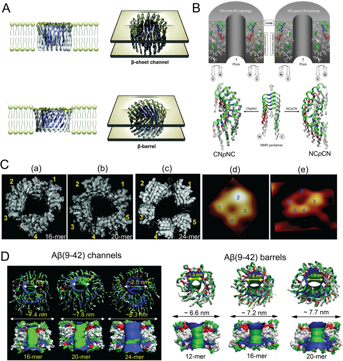
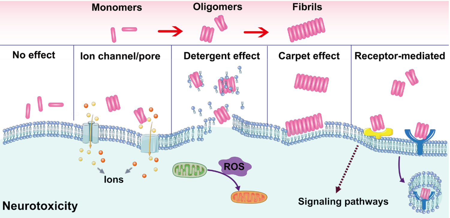
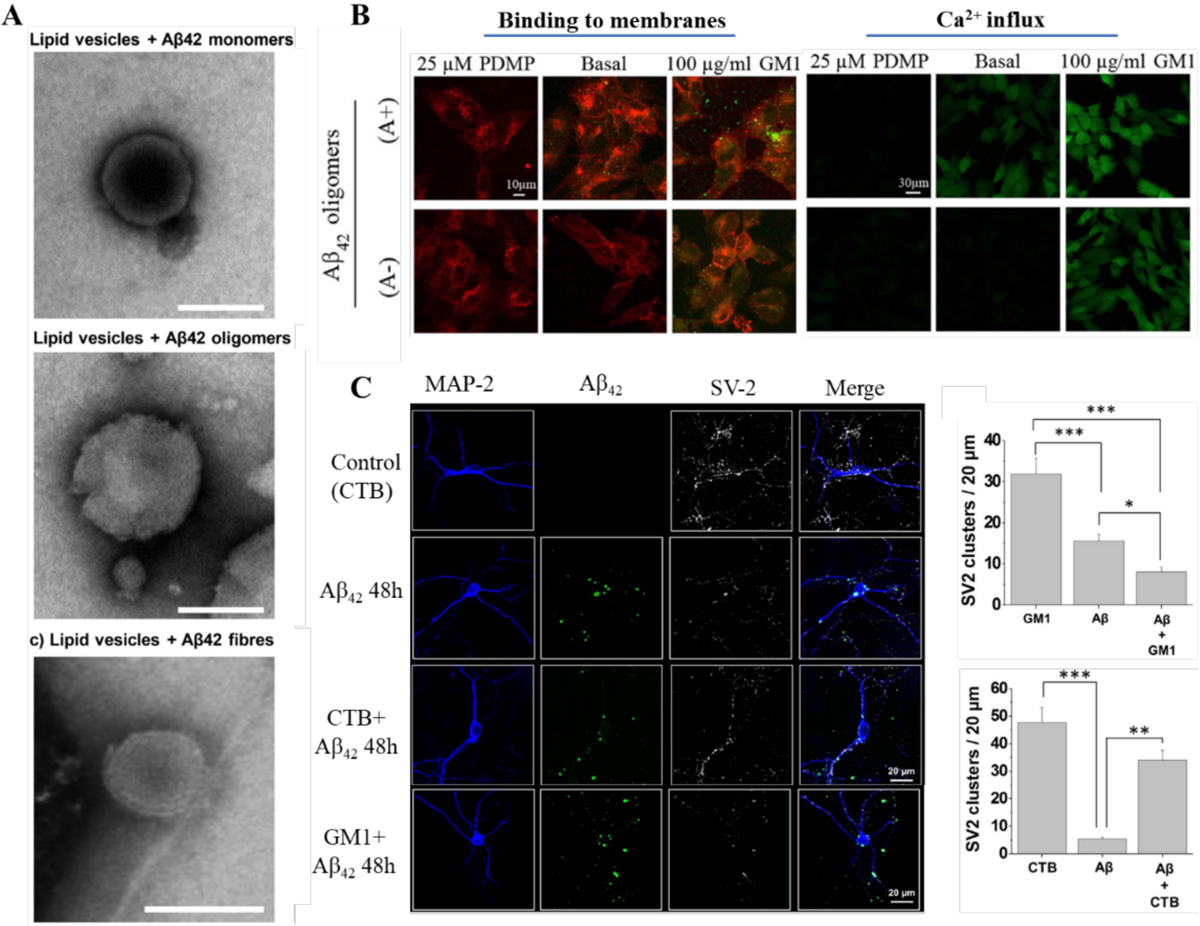
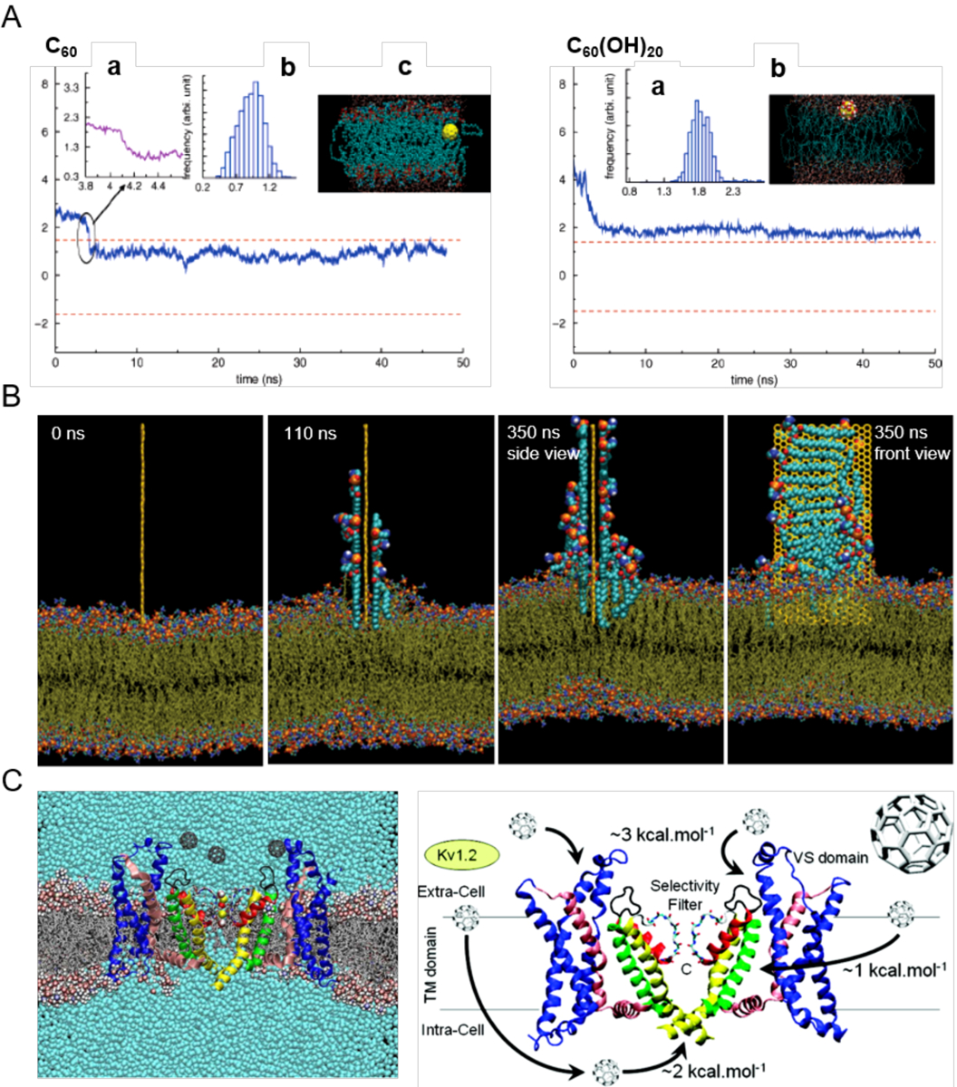
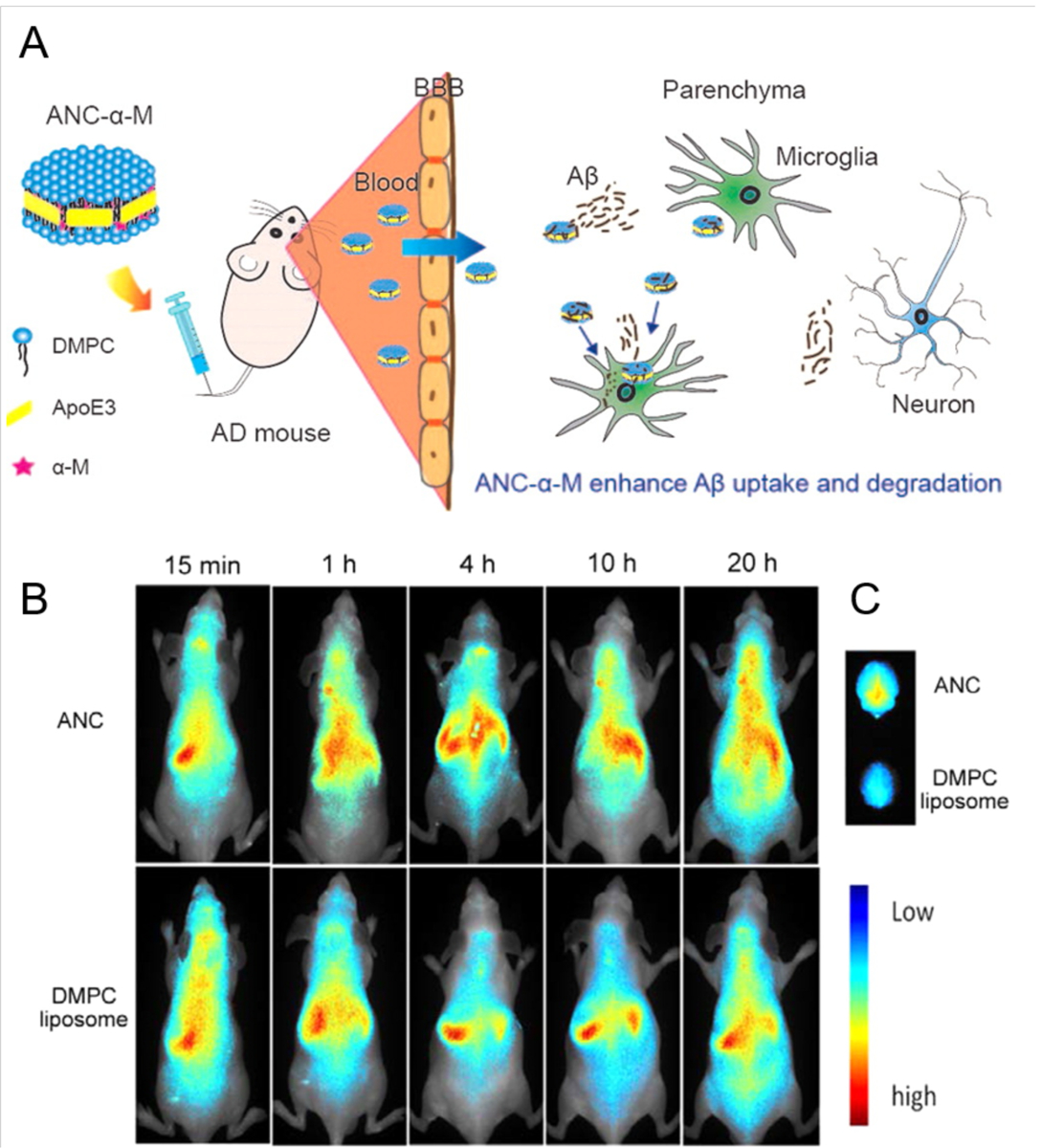
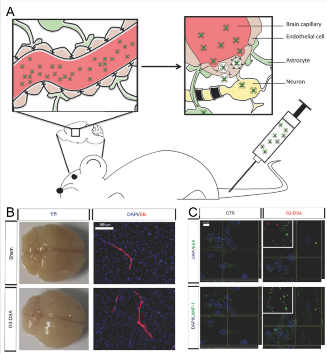
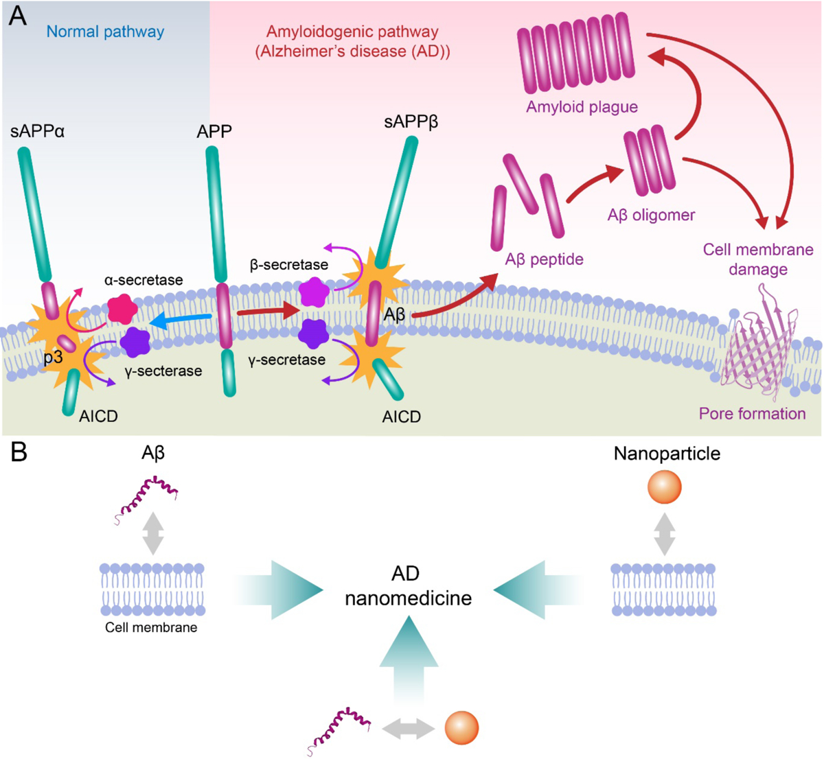
Alzheimer's disease (AD) is a major neurological disorder impairing its carrier's cognitive function, memory and lifespan. While the development of AD nanomedicine is still nascent, the field is evolving into a new scientific frontier driven by the diverse physicochemical properties and theranostic potential of nanomaterials and nanocomposites. Characteristic to the AD pathology is the deposition of amyloid plaques and tangles of amyloid beta (Aβ) and tau, whose aggregation kinetics may be curbed by nanoparticle inhibitors via sequence-specific targeting or nonspecific interactions with the amyloidogenic proteins. As literature implicates cell membrane as a culprit in AD pathogenesis, here we summarize the membrane axis of AD nanomedicine and present a new rationale that the field development may greatly benefit from harnessing our existing knowledge of Aβ-membrane interaction, nanoparticle-membrane interaction and Aβ-nanoparticle interaction.

Keywords: AD; Aβ; membrane; nanomedicine; nanoparticle.


Conflict of interest statement

Declaration of Competing Interest There are no conflicts to declare.

Figures

Figure 1.
Figure 1.. Aβ channels and β-barrels in lipid membranes.
(A) Schematic illustrations of Aβ channels and β-barrels in lipid membranes. (B) Schematics of the CNpNC (left) and NCpCN (right) topologies of Aβ(17–42) channels. The Aβ monomers, which were taken from the NMR pentamer structure in the PDB databank (ID: 2BEG), exhibited the U-shaped strand-turn-strand conformation. Top panel: initial annular channel topologies shown as a cross-section of a hollowed cylinder in grey with a cut along the pore axis. The Aβ(17–42) peptide in ribbon representation was projected into the cross-section area. Middle panel: the topologies of Aβ peptides drawn by connected arrows. Bottom panel: ribbon representations of Aβ backbones. (C) Comparison between computed Aβ(17–42) channel structures and high-resolution AFM data. The simulated channel structures for (a) 16-mer, (b) 20-mer and (c) 24-mer showed four to five subunits, in agreement with the AFM images (d and e). (D) Aβ(9–42) channels (left) and β-barrels (right). Top panel: angle views of the pore structures, where pore structures were shown in ribbon representation. Bottom panel: lateral views of the pore structure, where the cross-sectioned pores were shown in surface representation with the degree of the pore diameter colored in the order of red < green < blue. Panels reproduced from Refs. , –, –. Copyrights 2014 The Royal Society of Chemistry, 2007 the Biophysical Society, 2010 American Chemical Society, 2010 National Academy of Sciences and 2010 Elsevier Ltd.
Figure 2.
Figure 2.. Hypothesized mechanisms of Aβ-membrane interaction.
Carpet effect, ion channel, pore formation, detergent effect and receptor-mediated interaction are the most accepted modes of interaction.
Figure 3.
Figure 3.. Aβ-membrane interaction and subsequent biological effects.
(A) Disruption of lipid vesicles by Aβ42 species. (B) Binding of Aβ42 oligomers to cells with different GM1 contents and ensuing Ca2+ influxes. GM1-depleted (25 μM D-threo-1-phenyl-2-decanoylamino-3-morpholino-1-propanol, PDMP), basal and GM1 enriched (100 μ g/mL GM1) cells treated for 1 h with 10 μM A+ (toxic, obtained by 1 day incubation) or A (benign, obtained by 4 day incubation) Aβ42 oligomers. Red and green fluorescence emissions were from the cell membranes and Aβ42 oligomers, respectively. (C) Confocal micrographs and quantification of fluorescent Aβ clusters showing the distribution of Aβ clusters on the cell membrane surface along with the synaptic vesicle protein 2 (SV2) immunoreactivity in hippocampal neurons treated with Aβ-FAM. Cholera Toxin Subunit-B could block the interaction of Aβ with GM1. Panels reproduced from Refs. , –. Copyrights 2019 The American Society for Biochemistry and Molecular Biology, Inc., 2016 Springer Nature, 2017 Elsevier B.V.
Figure 4.
Figure 4.. Nanoparticle-membrane interactions in silico.
(A) Uptake of C60 (left) and C60(OH)20 (right) in the transmembrane (z) direction. The two dashed lines denote the upper and lower leaflets of the DPPC bilayer. Top left panel: (a) Zoomed trajectory at t = 4.09 ns. (b) Center-of-mass (COM) of C60 after it enters the bilayer (t > 4.2 ns). (c) Side view of the simulation system at t = 34.5 ns. The yellow ball denotes the C60 particle, cyan dots denote the lipid tail groups, and the red and blue dots represent the lipid head groups. Top right panel: (a) COM of C60(OH)20 during simulation. (b) Side view of the simulation system. Yellow balls and the attached large red and white dots denote the C60(OH)20 particle, cyan dots denote the tail groups of the DPPC lipids, and the small red and blue dots denote the lipid head groups.[97] (B) Lipid extraction by graphene in docking simulations over time. The restrained graphene nanosheet was docked at the surface of the outer POPE membrane of E. coli.[100] (C) Affinity of C60 with membrane potassium channels. (Left) A Kv1.2 channel embedded in a POPC lipid bilayer in the presence of C60 nanoparticles and their associated energetics.[101] Panels reproduced from Refs. , , . Copyrights 2007 American Chemical Society, 2013 Springer Nature and 2010 American Chemical Society.
Figure 5.
Figure 5.. Coarse-grained simulations of Aβ-nanoparticle interactions.
Dependence of Aβ aggregation on nanoparticle–protein interaction strength. (A) Average number of residues per chain that formed inter-peptide beta-sheets, Nβ–Res as a function of simulation time. (B) Maximum Nβ–Res as a function of nanoparticle-protein interaction strength εNP. Representative conformations with (C) εNP = 0.3ε (D) εNP = 0.74ε, where ε is the protein contact energy. (E) With a high protein/nanoparticle ratio, the nanoparticles (depicted as gray solid spheres) may promote the formation of amyloid fibrils (represented by red arrows) by increasing the local concentration of Aβ peptides on the nanoparticle surface (denoted as lines in cyan). (F) With a low protein/nanoparticle ratio, the nanoparticles displayed an inhibitive effect on the Aβ aggregation by depleting the peptides in solution but effectively having a low peptides concentration on nanoparticle surface. Reproduced with permission from Ref. . Copyrights 2015 The Royal Society of Chemistry.
Figure 6.
Figure 6.. Modes of action for nanoparticle inhibitors against Aβ amyloidosis.
Figure 7.
Figure 7.. Mitigation of Aβ toxicity and Alzheimer’s-like symptoms in adult zebrafish with βCas AuNPs.[24]
(A) Adult zebrafish (10 months old) were microinjected (cerebroventricular) with Aβ (1 μL, 50 μM). βCas AuNPs were microinjected (retro-orbital, 1 μL, 0.5 mM) 2 h prior to Aβ treatment. (B) Immunohistochemistry was performed on adult zebrafish brain sections to image the Aβ deposition. The first column represents the right cerebral brain of adult zebrafish in the GFP channel (Scale bars: 200 μM). DAPI, GFP and merged images at higher magnifications revealed Aβ plaque deposition in Aβ treated but not in Aβ + βCas AuNPs, or untreated control (Scale bars: 20 μM). (C) Cognitive behavior of adult zebrafish was analyzed. The movement trajectories of the fish in arena 1 vs. arena 2 are presented in the left panel. Comparative analysis of distance traveled and movement frequency of the fish in arena 1 vs. arena 2 revealed cognitive dysfunction of the Aβ-treated fish that were unable to avoid arena 2. Reproduced with permission from Ref. . Copyrights 2015 The Royal Society of Chemistry.
Figure 8.
Figure 8.. Nanocomposite-mediated neurotoxicity mitigation and Aβ removal in an AD mouse model.
(A) The Aβ-nanocomposite interaction entailed mitigation of Aβ aggregation and associated neurotoxicity. (B) Fluorescence images showing the Aβ/NC-KLVFF clusters elimination by BV-2 cells pretreated with or without TNF-α. Aβ was labeled with FITC (green), while NC-KLVFF was labeled with rhodamine B (red). Scale bar: 25 μm. (C) The nanocomposite mitigated the neurotoxicity elicited by Aβ aggregation in an AD mouse model. In situ neuronal apoptosis in hippocampal dentate gyrus subregion was labeled red. Reproduced with permission from Ref. . Copyrights 2019 American Chemical Society.
Figure 9.
Figure 9.. ApoE3-HDL nanomedicine for the mitigation of Aβ pathology.
(A) α-Mangostin loaded ApoE3-HDL nanocarriers (ANC-α-M) were able to translocate across the BBB of SAMP8 mice to promote Aβ clearance and rescue memory loss. (B) In vivo distribution in nude mice was studied after conjugating the ANC and DMPC liposomes, as control, with DIR fluorescence dye and administering to the mice via intravenous tail injection. (C) The ANCs were distributed to the brain with higher affinity, due to the presence of ApoE3. The figure is reproduced with permission from Ref. . Copyrights 2016 American Chemical Society.
Figure 10.
Figure 10.. Permeation of dendritic polymers across the BBB.
(A) Streptavidin-conjugated PAMAM dendrimers were able to permeate through the BBB via transcytosis in P21 mice. (B) Generation-3 DSA (succinamic acid dendrimer with curcumin) did not disrupt the BBB integrity. No extravasation of Evans blue (EB) dye was observed in whole brain epifluorescence, after administration of G3-DSA and Sham (positive control). (C) Endosomal trafficking of G2-DSA (generation-2 dendrimers with different length and charge of the dendron molecule) through bEnd.3 mouse neuronal cells. Colocalization of DSA positive vesicles (red) and early endosome antigen (EEA; green) for early endosomes and lysosomal-associated membrane protein (LAMP1; green) for late endosomes was observed. Figure is reproduced from Ref. , an open access publication from Advanced Science. Copyrights 2018 John Wiley & Sons.
Scheme.
Scheme.. Scope of the review.
(A) Aβ is synthesized in situ by the consecutive cleavage of transmembrane amyloid precursor protein (APP) by β and γ secretases. Under aberrant conditions Aβ peptides self-assemble to render oligomeric species and amyloid fibrils, which may compromise membrane integrity via various mechanisms, such as poration. (B) The development of AD nanomedicines may benefit from better understanding of Aβ-membrane, nanoparticle-membrane and Aβ-nanoparticle interactions. AICD: APP intracellular domain; sAPPβ: β-secretase cleaved soluble APP N-terminal fragment; sAPPα: α-secretase cleaved soluble extracellular APP fragment; p3: 3-kDa peptide resulting from α- and γ-secretase cleavage of APP.

Similar articles

Cited by

References

    1. Roher AE, Esh CL, Kokjohn TA, Castaño EM, Van Vickle GD, Kalback WM, Patton RL, Luehrs DC, Daugs ID, Kuo Y-M, Emmerling MR, Soares H, Quinn JF, Kaye J, Connor DJ, Silverberg NB, Adler CH, Seward JD, Beach TG, Sabbagh MN, Alzheimer’s & Dementia 2009, 5, 18. - PMC - PubMed
    1. Hardy J, Selkoe DJ, Science 2002, 297, 353. - PubMed
    1. Österlund N, Luo J, Wärmländer SKTS, Gräslund A, Biochim. Biophys. Acta Proteins Proteom 2019, 1867, 492. - PubMed
    1. Fabiani C, Antollini SS, Front. Cell. Neurosci 2019, 13. - PMC - PubMed
    1. Laurén J, Gimbel DA, Nygaard HB, Gilbert JW, Strittmatter SM, Nature 2009, 457, 1128. - PMC - PubMed