. 2020 Nov 25;1(1):ltaa001.
doi: 10.1093/immadv/ltaa001. eCollection 2021 Jan.

Protective low-avidity anti-tumour CD8+ T cells are selectively attenuated by regulatory T cells

Affiliations

Protective low-avidity anti-tumour CD8+ T cells are selectively attenuated by regulatory T cells

Gessa Sugiyarto et al. Immunother Adv. .

Abstract

Objectives: Regulatory T cells (Treg) play a major role in the suppression of protective anti-tumour T cell responses. In the CT26 BALB/c murine model of colorectal carcinoma, Tregs differentially suppress responses to two characterised CD8+ T epitopes, AH1 and GSW11, which results in an absence of detectable IFN-γ-producing GSW11-specific T cells in the spleen and lymph nodes of tumour challenged mice. Activation of GSW11-specific T cells correlates with protection against tumour progression. We wanted to examine the presence of non-functional GSW11-specific T cells in Treg replete and depleted mice, assess their phenotype and their affinity compared to AH1-specific T cells.

Methods: We used peptide-specific tetramers to identify tumour-specific CD8+ T cells and assessed the cell surface expression of markers associated with exhaustion (PD-1, Tim3 and Lag-3) and their function by IFN-g production using flow cytometry. We also assessed the T cell receptor (TcR) clonality of tumour-specific T cells. Tetramer competition assays were performed to determine the relative affinity of identified TcR.

Results: Here, we show that GSW11-specific T cells are in fact induced in Treg-replete, CT26-bearing mice, where they make up the majority of tumour-infiltrating CD8+ lymphocytes, but exhibit an 'exhausted' phenotype. This dysfunctional phenotype is induced early in the anti-tumour response in tumours. Depletion of Tregs prior to tumour challenge correlates with an altered T cell receptor (TcR) repertoire. Moreover, the avidity of GSW11-specific TcRs that expanded in the absence of Tregs was significantly lower compared with TcRs of CD8+populations that were diminished in protective anti-tumour responses.

Conclusion: Our results indicate that Tregs suppress the induction of protective anti-tumour T cell responses and may signify that low-avidity T cells play an important role in this protection.

Keywords: antigen presentation; immunotherapy; regulatory T cell; tumour immunity.


Figures

Graphical Abstract
Graphical Abstract
Figure 1.
Figure 1.
The majority of tumour-infiltrating GSW11-specific T cells are non-functional. BALB/c mice were challenged with CT26 tumour cells and the presence of tumour-infiltrating AH1- and GSW11-specific T cells was assessed over the indicated time course. (A) Assessment of AH1- and GSW11-specific T cells using tetramer/dextramer and IFN-γ production at day 10 and day 22. (B) Percentage of antigen-specific T cells detected by tetramer/dextramer. (C) Diagram representing the tumour size (diameter) and relative proportions of antigen-specific tumour-infiltrating T cells over time. (D) Percentage of functional AH1- and GSW11-specific CD8+ T cells. (E) Relative proportion of functional and non-functional antigen-specific tumour-infiltrating T cells. (B, D; mean and SEM of two/three mice at each time point; *P < 0.05, ***P < 0.001).
Figure 2.
Figure 2.
Tumour-infiltrating GSW11-specific T cells are more exhaustible than AH1-specific T cells. (A) A representative gating strategy to assess Lag-3 and Tim3 expression on PD-1int and PD-1hi CD8+ T cells. BALB/c mice were challenged with CT26 tumour and the number of antigen-specific CD8+ T cells/100,000 cells was assessed at early (day 7) and late (day 22) stages of tumour progression was assessed (B). The number of tumour-infiltrating PD-1hi, Lag3+, Tim-3+ GSW11-, and AH1-specific CD8+ T cells (C) and the number of those cells that produced IFN-γ following peptide antigen stimulation was evaluated (D). Assessment of the number of functional tumour-infiltrating PD-1int GSW11- and AH1-specific CD8+ T cells (E). B–E; mean and SEM of three mice at each time point; *P < 0.05, **P < 0.01).
Figure 3.
Figure 3.
Draining lymph node tumour-specific CD8+ T cells become exhausted as tumour progresses. BALB/c mice were challenged with CT26 tumour and antigen-specific CD8+ T cells in tumour draining lymph nodes were characterised. (A) A representative histogram showing PD-1 expression on naive GSW11- and AH1-specific CD8+ T cells. (B) The number of antigen-specific CD8+ T cells present in tumour draining lymph nodes during tumour progression. (C) The percentage of functional PD-1int GSW11- and AH1-specific CD8+ T cells in draining lymph nodes. Assessment of the percentage of PD-1hi, Lag3+, Tim-3+ GSW11- and AH1-specific CD8+ T cells (D) and the percentage that produced IFN-γ following antigen stimulation (E) in tumour draining lymph nodes. (B–E; mean and SEM of three mice at the indicated time points).
Figure 4.
Figure 4.
GSW11-specific T cell clonalities are modulated by regulatory T cells. Treg replete or depleted BALB/c mice were challenged with CT26 and the TcR expression of GSW11-specific T cells assessed at day 7. (A) Percentage of GSW11-specific CD8+ T cells expressing the indicating TcR Vβ in Treg replete or depleted CT26 challenged mice. The percentage of exhausted PD-1hi, Tim3+, Lag-3+ GSW11-specific CD8+ T cells expressing TcR unchanged, increased or decreased in Treg depleted mice as individual TcR (B) or grouped (C). (A; mean and SEM of 10 mice in three pools, B; mean of ten mice from three pools, C and D; mean of at least three mice from two independent experiments; *P < 0.05, **P < 0.01, ***P < 0.001, ****P < 0.0001).
Figure 5.
Figure 5.
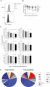
Tumour protective GSW11-specific CD8+ T cells have low TcR avidity. The TcR avidity of anti-GSW11 T cell oligoclones that showed either increased, decreased, or similar levels in tumour challenged Treg depleted mice was assessed using tetramer competition. (A, B) A representative histogram (A) and analysis of relative change in tetramer MFI (B) of the TcR tetramer competition assay in indicated TcR clones. (C) Relative avidity of indicated GSW11-specific T cell oligoclones following tetramer competition. (D) The relative proportion of TcR Vβ usage in GSW11-specific T cells following tumour challenge (blue segments indicate TcR with high avidity and red indicate those with low avidity). (C; mean and SEM of at least three independent experiments and D; mean of at least three mice from two independent experiments; *P < 0.05, **P < 0.01, ****P < 0.0001).

References

    1. Hiraoka K, Miyamoto M, Cho Y et al. Concurrent infiltration by CD8+ T cells and CD4+ T cells is a favourable prognostic factor in non-small-cell lung carcinoma. Br J Cancer 2006;94(2):275–80. doi:10.1038/sj.bjc.6602934 - DOI - PMC - PubMed
    1. Naito Y, Saito K, Shiiba K et al. CD8+ T cells infiltrated within cancer cell nests as a prognostic factor in human colorectal cancer. Cancer Res 1998;58(16):3491–4. - PubMed
    1. Nakano O, Sato M, Naito Y et al. Proliferative activity of intratumoral CD8(+) T-lymphocytes as a prognostic factor in human renal cell carcinoma: clinicopathologic demonstration of antitumor immunity. Cancer Res 2001;61(13):5132–6. - PubMed
    1. Pages F, Berger A, Camus M et al. . Effector memory T cells, early metastasis, and survival in colorectal cancer. N Engl J Med 2005;353(25):2654–66. doi:10.1056/NEJMoa051424 - DOI - PubMed
    1. Sato E, Olson SH, Ahn J et al. Intraepithelial CD8+ tumor-infiltrating lymphocytes and a high CD8+/regulatory T cell ratio are associated with favorable prognosis in ovarian cancer. Proc Natl Acad Sci USA 2005;102(51):18538–43. doi:10.1073/pnas.0509182102 - DOI - PMC - PubMed