. 2021 Aug;86(2):791-803.
doi: 10.1002/mrm.28748. Epub 2021 Mar 10.

Distortion-free, high-isotropic-resolution diffusion MRI with gSlider BUDA-EPI and multicoil dynamic B0 shimming

Affiliations

Distortion-free, high-isotropic-resolution diffusion MRI with gSlider BUDA-EPI and multicoil dynamic B0 shimming

Congyu Liao et al. Magn Reson Med. 2021 Aug.

Abstract

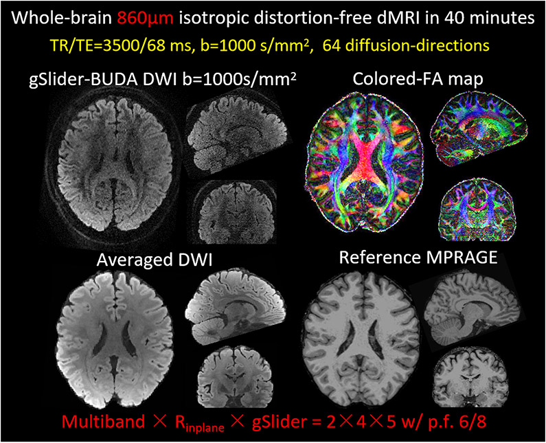
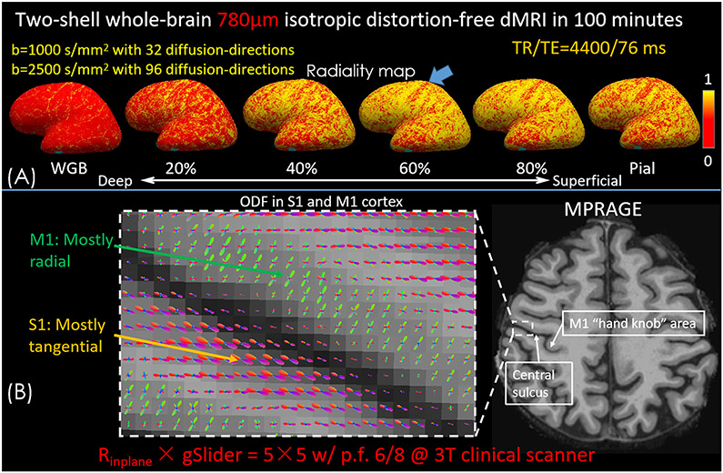
Purpose: We combine SNR-efficient acquisition and model-based reconstruction strategies with newly available hardware instrumentation to achieve distortion-free in vivo diffusion MRI of the brain at submillimeter-isotropic resolution with high fidelity and sensitivity on a clinical 3T scanner.

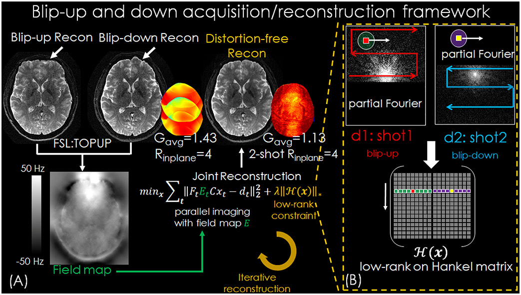
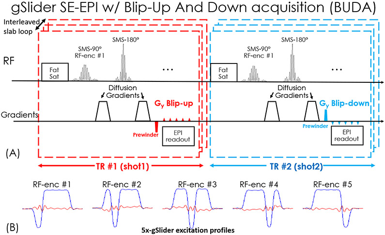
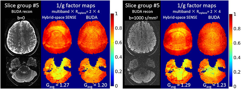
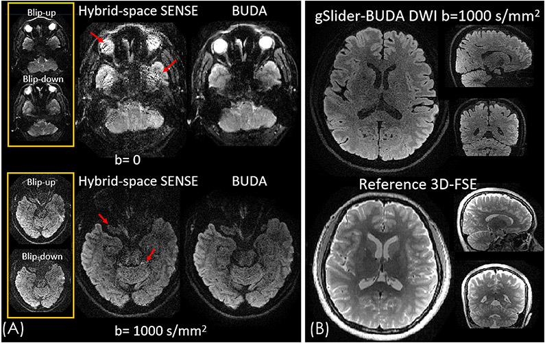
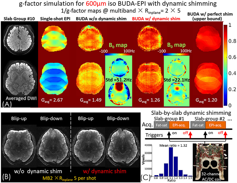
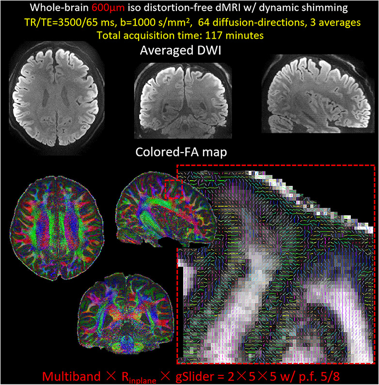
Methods: We propose blip-up/down acquisition (BUDA) for multishot EPI using interleaved blip-up/blip-down phase encoding and incorporate B0 forward-modeling into structured low-rank reconstruction to enable distortion-free and navigator-free diffusion MRI. We further combine BUDA-EPI with an SNR-efficient simultaneous multislab acquisition (generalized slice-dithered enhanced resolution ["gSlider"]), to achieve high-isotropic-resolution diffusion MRI. To validate gSlider BUDA-EPI, whole-brain diffusion data at 860-μm and 780-μm data sets were acquired. Finally, to improve the conditioning and minimize noise penalty in BUDA reconstruction at very high resolutions where B0 inhomogeneity can have a detrimental effect, the level of B0 inhomogeneity was reduced by incorporating slab-by-slab dynamic shimming with a 32-channel AC/DC coil into the acquisition. Whole-brain 600-μm diffusion data were then acquired with this combined approach of gSlider BUDA-EPI with dynamic shimming.

Results: The results of 860-μm and 780-μm datasets show high geometry fidelity with gSlider BUDA-EPI. With dynamic shimming, the BUDA reconstruction's noise penalty was further alleviated. This enables whole-brain 600-μm isotropic resolution diffusion imaging with high image quality.

Conclusions: The gSlider BUDA-EPI method enables high-quality, distortion-free diffusion imaging across the whole brain at submillimeter resolution, where the use of multicoil dynamic B0 shimming further improves reconstruction performance, which can be particularly useful at very high resolutions.

Keywords: diffusion-weighted imaging; distortion correction; gSlider; high-isotropic resolution; shim array.


Figures

Figure 1.
Figure 1.
(A) Reconstruction flowchart of BUDA-EPI. (B) Complementary k-space acquisition strategy is applied to each shot and the reconstructed multi-shot data are reshaped to the block-Hankel representation and enforced by the low rank prior in each iteration to provide robustness to shot-to-shot phase corruption.
Figure 2.
Figure 2.
(A) Sequence diagram of gSlider BUDA-EPI. (B) The designed 5x-gSlider excitation profiles.
Figure 3.
Figure 3.
The reconstruction results and 1/g-factor maps of the b=0 and 1000 s/mm2 EPI data, with MB × Rinplane = 2 × 4 accelerations.
Figure 4.
Figure 4.
(A) Comparison between hybrid-space SENSE and BUDA reconstructions for an acquisition at MB × Rinplane = 2 × 4 (B) The diffusion weighted volume reconstructed by gSlider-BUDA method. The diffusion weighted volume retains high geometric fidelity that closely resembled that of the reference 3D-FSE images.
Figure 5.
Figure 5.
Three orthogonal views of i) whole-brain single-direction diffusion-weighted images (DWI), ii) colored-FA maps and iii) 64-diffusion-direction averaged DWI from gSlider BUDA-EPI at 860μm isotropic resolution. As can be seen from the averaged DWI volume, the diffusion data retains high geometric fidelity with the reference MPRAGE.
Figure 6.
Figure 6.
(A) Radiality maps at the surface of the white-gray boundary (WGB) surface, the surfaces of 20%, 40%, 60% and 80% of cortical thickness into gray matter, and the pial surface on the inflated brain surface. (B) ODF results in primary motor cortex (M1) and somatosensory cortex (S1) obtained from the whole brain 780μm dMRI data with two shells.
Figure 7.
Figure 7.
(A) 1/g-factor maps of single-shot EPI, BUDA-EPI without and with dynamic shim, and with perfect shim (no B0 inhomogeneity); all at MB × Rinplane= 2 × 5. (B) Using the 32-channel AC/DC coil with slab-by-slab dynamic shimming, the distortion difference between blip-up and blip-down shots are mitigated and hence (C) the average g-factor of BUDA reconstruction improves by 32% compared to no dynamic shimming.
Figure 8.
Figure 8.
The averaged diffusion-weighted images and the colored-FA maps from 64-diffusion-direction whole-brain 600μm gSlider BUDA-EPI with dynamic shimming. The zoom-in sagittal view of a slice of colored FA maps, displaying the high-resolution capability of the 600μm data.

Similar articles

Cited by

References

    1. Leuze CWU, Anwander A, Bazin PL, et al. Layer-specific intracortical connectivity revealed with diffusion MRI. Cereb. Cortex 2014;24:328–339 doi: 10.1093/cercor/bhs311. - DOI - PMC - PubMed
    1. Assaf Y Imaging laminar structures in the gray matter with diffusion MRI. Neuroimage 2019;197:677–688 doi: 10.1016/j.neuroimage.2017.12.096. - DOI - PubMed
    1. Ganepola T, Nagy Z, Ghosh A, Papadopoulo T, Alexander DC, Sereno MI. Using diffusion MRI to discriminate areas of cortical grey matter. Neuroimage 2018;182:456–468 doi: 10.1016/j.neuroimage.2017.12.046. - DOI - PMC - PubMed
    1. Song AW, Chang HC, Petty C, Guidon A, Chen NK. Improved delineation of short cortical association fibers and gray/white matter boundary using whole-brain three-dimensional diffusion tensor imaging at submillimeter spatial resolution. Brain Connect. 2014;4:636–640 doi: 10.1089/brain.2014.0270. - DOI - PMC - PubMed
    1. Bianciardi M, Toschi N, Edlow BL, et al. Toward an In Vivo Neuroimaging Template of Human Brainstem Nuclei of the Ascending Arousal, Autonomic, and Motor Systems. Brain Connect. 2015;5:597–607 doi: 10.1089/brain.2015.0347. - DOI - PMC - PubMed

Publication types

MeSH terms