. 2021 May;69(5):321-338.
doi: 10.1369/00221554211004297. Epub 2021 Mar 22.

Large- and Medium-sized Arteries Remaining in Transmural Scar Distal to Permanent Coronary Ligation Undergo Neointimal Hyperplasia and Inward Remodeling

Affiliations

Large- and Medium-sized Arteries Remaining in Transmural Scar Distal to Permanent Coronary Ligation Undergo Neointimal Hyperplasia and Inward Remodeling

Eduard I Dedkov. J Histochem Cytochem. 2021 May.

Abstract

This study aimed to investigate the structural integrity and dynamic changes in chronically occluded residual arteries found in post-myocardial infarction (MI) scar. A transmural MI was induced in middle-aged, male Sprague-Dawley rats by left coronary artery ligation. The rats were euthanized 3 days and 1, 2, 4, 8, and 12 weeks after MI, and their hearts were processed into paraffin for histology, immunohistochemistry, and quantitative morphometry. It has been found that large- and medium-sized arteries were able to survive inside the transmural scars for 12 post-MI weeks. Furthermore, most residual arteries preserved their structural integrity for up to 2 weeks post-MI, but gradually all disused vessels had undergone neointimal hyperplasia and inward remodeling at later time periods. In addition, the replacement of vascular smooth muscle cells in the wall of residual arteries by extracellular matrix components led to a disruption of the vessel integrity and progressive obliteration of their lumen between 4 and 12 post-MI weeks. Taken together, this study demonstrate that residual arteries in post-infarcted region were capable of maintaining their structural integrity, including the patent lumen, during two post-MI weeks, suggesting that during this period they can be used as potential conduits for conceivable reflow of arterial blood within the scarred region of the heart.

Keywords: inward arterial remodeling; middle-aged rats; myocardial infarction; neointimal hyperplasia; residual coronary arteries; transmural scar.


Conflict of interest statement

Competing Interests: The author(s) declared no potential conflicts of interest with respect to the research, authorship, and/or publication of this article.

Figures

Figure 1.
Figure 1.
Residual coronary arteries in developing and mature transmural scars. The hematoxylin and eosin–stained micrographs demonstrate the profiles of surviving large- and medium-sized arteries in 3-day-old (C, D), 1-week-old (E, F), 2-week-old (G, H), 4-week-old (I, J), 8-week-old (K, L), and 12-week-old (M, N) post-MI scars. Note that the coronary artery shown in micrographs A and B serves as a representative example of a large arterial vessel in structurally intact LV myocardium of a sham-operated rat. In each low-power view (A, C, E, G, I, K, and M), an arrow points to a rectangular outline designating a location of the corresponding coronary arteries which are displayed in high-power micrographs B, D, F, H, J, L, and N, respectively. The arrowheads in micrographs C, E, G, I, K, and M mark the edges of the transmural scar. The asterisks in micrographs D, F, and H identify the vascularized granulation tissue, whereas in micrographs J, L, and M, they denote the fibrous tissue which surrounds the residual coronary arteries in the scars. Scale bars are 4 mm (C, G, I, and K), 3 mm (A, E, and M), 100 µm (B, D, F, and H), and 50 µm (J, L and N). Abbreviations: D, days; WK, week(s); MI, myocardial infarction.
Figure 2.
Figure 2.
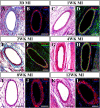
Expression of muscle-specific proteins in vascular smooth muscle (VSM) cells of the residual coronary arteries in transmural scars 3 days (A, B), 1 week (C, D), 2 weeks (E, F), 4 weeks (G, H), 8 weeks (I, J), and 12 weeks (K, L) after MI. Note, in each time point, the two micrographs demonstrate the same arterial vessel found on adjacent serial sections which were stained either with histological dyes or with immunofluorescence technique. A combination between these two staining methods has allowed to confirm that the vessels demonstrating the expression of muscle-specific protein, such as α-smooth muscle actin (α-SMA) and desmin, were the same which were initially identified on the sections stained with histological dyes only. The histological stains used to visualize the residual coronary arteries in the scars were Masson’s trichrome, in A and E, and H&E, in C, G, I, and K. The corresponding serial sections were immunofluorescence stained either with an antibody against α-SMA (red color in B, J, and L) or with a combination of the antibodies against desmin (red color) and laminin (green color) in D, F, and H. In the latter, the presence of laminin-positive labeling beneath the endothelial layer and around VSM cells confirms the maintenance of the basement membranes in the wall of the residual arteries. All immunofluorescence-labeled sections were counterstained with DAPI (blue color) to visualize the nuclei. Scale bars are 100 µm (A, B, C, D, E, F, F, H, J, and K) and 50 µm (C, I, and L). Abbreviations: D, days; WK, week(s); H&E, hematoxylin and eosin; α-SMA, α-smooth muscle actin; DAPI, 4′, 6-diamino-2-phenylindole; MI, myocardial infarction.
Figure 3.
Figure 3.
Degenerative changes observed in coronary arteries remaining in transmural scars during two post-MI weeks. (A) An H&E-stained section of a 3-day-old post-MI scar demonstrating hyaline degeneration in the tunica media of a small-diameter arterial branch (arrowheads) that remains in alignment with a surviving portion of the large residual coronary artery. Note a massive accumulation of extravasated erythrocytes surrounding the degenerating arterial branch. The arrows denote the point of transition between necrotic and surviving segments in the artery. (B, C) and (D, E) Two representative necrotic coronary arteries identified in serial sections of a 1-week-old post-MI scar stained with H&E, in B and D, and with Masson’s trichrome, in C and E, respectively. Note that the branches of the same arterial vessel shown in B and C display the features of fibrinoid necrosis associated with leukocytic infiltration (arrows) and accumulation of fibrin-like material (asterisks) in the vessel wall, whereas the large artery depicted in D and E reveals the characteristic manifestations of hyaline degeneration associated with the homogeneously stained, acellular tunica media and a well-preserved endothelial lining (arrowheads). (F, G) and (H, I) Two representative necrotic coronary arteries recognized in serial sections of a 2-week-old post-MI scar stained with H&E, in F and H, and with Masson’s trichrome, in G and I, respectively. Note that the same arterial vessel shown in F and G displays the signs resembling fibrinoid necrosis (asterisks), whereas the artery illustrated in H and I shows the typical appearance of hyaline degeneration of the tunica media. The arrowheads in H and I point to the nuclei of surviving endothelial cells. (J, K) The same representative region from a 2-week-old post-MI scar stained with H&E (J) and Masson’s trichrome (K) demonstrating the presence of a small arterial vessel with the features of fibrinoid necrosis (asterisk) in close proximity to a structurally intact larger artery (arrows). All scale bars are 100 µm. Abbreviations: D, days; WK, week(s); H&E, hematoxylin and eosin; MI, myocardial infarction.
Figure 4.
Figure 4.
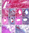
Local variability in the degree of structural alterations among the residual arteries in 1-week-old (A–C) and 2-week old (D–F) post-MI scars. Note that each individual set of the micrographs (A–C or D–F) displays the profiles of the same representative arteries identified in adjacent serial sections stained with H&E (A, D), Masson’s trichrome (B, E), and picrosirius red (C, F). In each micrograph, the asterisk denotes an artery that demonstrates more advanced structural changes, such as the thickening of the arterial wall and accumulation of the extracellular matrix, including collagen fibers, in the tunica media. Scale bars are 100 µm. Abbreviations: WK, week(s); H&E, hematoxylin and eosin; MI, myocardial infarction.
Figure 5.
Figure 5.
The pattern of structural modifications detected in coronary arteries surviving in 1-week-old (A–H) and 2-week-old (I–P) post-MI scars. Note that each individual set of the micrographs (A–D or I–L) demonstrates the same representative artery recognized on adjacent serial sections stained with H&E (A, I), Masson’s trichrome (B, J), and picrosirius red (C, K) and with an antibody against α-SMA that was visualized using 3, 3′-diaminobenzidine chromogen (brown color in D, L). In all light-microscopy images, the black, dotted line delineates the border between the tunica media and neointimal hyperplasia. A combination of different stains applied to serial profiles of the same arteries has confirmed that most of the cells forming the neointima in these vessels were activated or synthetic vascular smooth muscle (VSM) cells (D and L). A switch from a contractile to a synthetic phenotype in these cells is supported by the absence of a spindle-shaped morphology (A, B and I, J) and progressive accumulation of collagen fibers around the individual cells (C and K). In addition, two sets of fluorescence micrographs (E–H and M–P) obtained from 1- and 2-week-old post-MI scar, respectively, display the representative residual coronary arteries immunostained with the antibodies against BrdU (green color in G, H and O, P) and α-SMA (red color in E–H and M–P) in a combination with the nuclear counterstain by DAPI (blue color in E, F and M, N). Note that micrographs F, H, N, and P represent the high-power views of the areas outlined by a box in micrographs E, G, M, and O, respectively. The nature of the growing (BrdU-labeled) cells in the neointima was established on the basis of two principal criteria: (1) the proximity of a BrdU-positive nucleus to the vessel lumen and (2) the presence or absence of α-SMA immunostaining in the cytoplasm. Such methodological approach has allowed to confirm that the arrowheads in F and H denote BrdU-positive/α-SMA-positive VSM cells within the neointima, whereas the arrows in N and P point to BrdU-positive/α-SMA-negative cells in the endothelial lining. To facilitate the tracing of all BrdU-labeled cells in corresponding images of the same arterial wall, each arrow or arrowhead in micrographs E, G or M, O points to the matching nucleus. According to these observations, the neointimal growth in the residual coronary arteries involved proliferation of both synthetic VSM cells (arrowheads in E, G and M, O) and the cells of the endothelial lining (arrowheads in E, G and M, O). Scale bars are 50 µm (A–D, E, G, I–L, M, and O) and 25 µm (F, H, N, and P). Abbreviations: WK, week(s); H&E, hematoxylin and eosin; α-SMA, α-smooth muscle actin; DAPI, 4′, 6-diamino-2-phenylindole; BrdU, 5-bromo-2′-deoxyuridine; MI, myocardial infarction.
Figure 6.
Figure 6.
The pattern of structural alterations observed in coronary arteries surviving in 4-week-old (A–D), 8-week-old (E–H), and 12-week-old (I–L) post-MI scars. Note that each individual set of the micrographs (A–D, E–H, or I–L) displays the same representative artery identified on adjacent serial sections stained with Masson’s trichrome (A, E), H&E (I), Verhoeff’s elastic tissue stain (C, G, and K), and picrosirius red (D, H, and L) and with an antibody against α-SMA that was visualized with 3, 3′-diaminobenzidine chromogen (brown color in B, F, and J). The white or black dotted outline seen on the arterial profiles in A, B, D, E, F, H, I, J, and L delineates the border between the tunica media and the neointima. In micrographs C, G, and K, the border between the tunica media and neointimal hyperplasia is demarcated by the internal elastic lamina visualized by Verhoeff’s elastic tissue stain as a black circumferential outline. Note that the discontinuous staining pattern of the internal elastic lamina seen in micrographs G and K (arrows) suggests some disruption of structural integrity in this essential component of the arterial wall. Furthermore, a combination of different stains applied to serial profiles of the same residual arteries has revealed that synthetic VSM cells of the neointima continued to deposit the amorphous and fibrous components of the extracellular matrix (A and E), including elastin (C, G, and K) and collagen fibers (D, H, and L), thereby thickening the arterial wall. However, it is important to highlight that despite substantial neointimal expansion, most surviving arteries were able to maintain a narrowed, but a patent, lumen. Scale bars are 50 µm. Abbreviations: WK, weeks; H&E, hematoxylin and eosin; α-SMA, α-smooth muscle actin; MI, myocardial infarction; VSM, vascular smooth muscle.
Figure 7.
Figure 7.
Time course of changes in external (A) and lumen (B) diameters and total wall thickness (C) and the total wall thickness-to-lumen ratio (D) in the residual coronary arteries remaining in transmural scars during 12 post-MI weeks. Note, the pattern of progressive structural modifications observed in a majority of the residual arteries resembles that of inward arterial remodeling characterized by a marked increase in the wall-to-lumen ratio (D) caused by prominent thickening of the vessel wall in a combination with a reduction of external (outer) and lumen (inner) vessel diameters. Data are mean ± SEM. Abbreviations: MI, myocardial infarction; SEM, standard error of the mean. *p≤0.05, **p≤0.01, ***p≤0.001 vs sham-operated (SH) rats; §p≤0.05, §§p≤0.01, §§§p≤0.001 vs 3-day (3D) post-MI rats; p≤0.05, ††p≤0.01, †††p≤0.001 vs 1-week (1W) post-MI rats; ‡‡‡p≤0.001 vs 2-week (2W) post-MI rats; #p≤0.05 vs 4-week (4W) post-MI rats.
Figure 8.
Figure 8.
Detrimental structural changes detected in some residual coronary arteries remaining in 4-week-old (A–D), 8-week-old (E–H), and 12-week-old (I, J and K, L) post-MI scars. Note that each individual set of the micrographs (A–D or E–H) demonstrates the same representative artery identified on adjacent serial sections stained with Masson’s trichrome (A, E), Verhoeff’s elastic tissue stain (C, G), and picrosirius red (D, H) and with an antibody against α-SMA that was visualized with 3, 3′-diaminobenzidine chromogen (brown color in B, F). The white or black dotted outline seen on the arterial profiles in E, F, and H defines the border between the tunica media and the neointima. In micrographs C and G, the border between the tunica media and neointimal hyperplasia is delineated by the internal elastic lamina visualized by Verhoeff’s elastic tissue stain as a black circumferential outline. A combination of different stains applied to serial profiles of the same residual arteries has revealed a patchy loss of VSM cells in both the tunica media (corresponding arrowheads in A, B and E, F) and the neointimal formation (corresponding double-headed arrows in A–D and an asterisk in E–H). Note that the areas which were formerly occupied by either contractile or synthetic VSM cells become filled with the extracellular material (A and E), including elastin (C and G) and collagen fibers (D and H). The analogous sporadic pattern of VSM cell disappearance has been also detected in the wall of some residual arteries from 12-week-old post-MI scars (I, J and K, L). In the latter micrographs, two representative arterial vessels were immunostained with the antibodies against desmin (red color in I–L) and laminin (green color in J and L) in a combination with the nuclear counterstain by DAPI (blue color in I–L). The laminin immunostaining was utilized to visualize the basement membranes of the VSM cells throughout the entire arterial wall. The patchy absence of desmin-positive immunostaining within the laminin-outlined arterial wall (asterisks in I–L) evidently confirms that these areas lack the VSM cells. Furthermore, it is important to emphasize that, in some residual vessels, the neointimal formation continued to accumulate the extracellular material around the remaining VSM cells (E), causing fibrosis of the arterial wall and marked narrowing of the vessel lumen. Moreover, the fragmented staining pattern of the internal elastic lamina in such vessels (arrows in C and G) suggests a complete loss of structural integrity in the arterial wall. Scale bars are 50 µm. Abbreviations: WK, weeks; α-SMA, α-smooth muscle actin; MI, myocardial infarction; VSM, vascular smooth muscle; DAPI, 4′, 6-diamino-2-phenylindole.

Similar articles

Cited by

References

    1. Groenewegen A, Rutten FH, Mosterd A, Hoes AW. Epidemiology of heart failure. Eur J Heart Fail. 2020;22:1342–56. doi:10.1002/ejhf.1858. - DOI - PMC - PubMed
    1. Pfeffer MA, Braunwald E. Ventricular remodeling after myocardial infarction. Experimental observations and clinical implications. Circulation. 1990;81(4):1161–72. - PubMed
    1. Bhatt AS, Ambrosy AP, Velazquez EJ. Adverse remodeling and reverse remodeling after myocardial infarction. Curr Cardiol Rep. 2017;19(8):71. doi:10.1007/s11886-017. - DOI - PubMed
    1. Yancy CW, Jessup M, Bozkurt B, Butler J, Casey DE, Jr, Colvin MM, Drazner MH, Filippatos GS, Fonarow GC, Givertz MM, Hollenberg SM, Lindenfeld J, Masoudi FA, McBride PE, Peterson PN, Stevenson LW, Westlake C. 2017 ACC/AHA/HFSA focused update of the 2013 ACCF/AHA guideline for the management of heart failure: a report of the American College of Cardiology/American Heart Association Task Force on Clinical Practice Guidelines and the Heart Failure Society of America. J Am Coll Cardiol. 2017;70(6):776–803. doi:10.1016/j.jacc.2017.04.025. - DOI - PubMed
    1. Sanganalmath SK, Bolli R. Cell therapy for heart failure: a comprehensive overview of experimental and clinical studies, current challenges, and future directions. Circ Res. 2013;113(6):810–34. doi:10.1161/CIRCRESAHA.113.300219. - DOI - PMC - PubMed